Loading
PDBj
MenuPDBj@FacebookPDBj@TwitterPDBj@YouTubewwPDB FoundationwwPDB
RCSB PDBPDBeBMRBAdv. SearchSearch help
Search by PDB author
8HSF
DownloadVisualize
BU of 8hsf by Molmil
Insulin triple mutant INS-RQD
Descriptor: Insulin A chain, Insulin B chain
Authors:Kundapura, S.V, Rao, S.S, Kulal, A, Ramagopal, U.A.
Deposit date:2022-12-19
Release date:2023-12-20
Method:X-RAY DIFFRACTION (2.9 Å)
Cite:Insulin triple mutant INS-RQD
To Be Published
7FDS
DownloadVisualize
BU of 7fds by Molmil
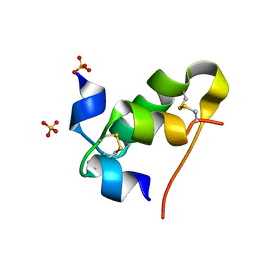
High resolution crystal structure of LpqH from Mycobacterium tuberculosis
Descriptor: Lipoprotein LpqH
Authors:Kundapura, S.V, Chatterjee, S, Samanta, D, Ramagopal, U.A.
Deposit date:2021-07-17
Release date:2021-12-15
Last modified:2023-11-29
Method:X-RAY DIFFRACTION (1.258 Å)
Cite:High-resolution crystal structure of LpqH, an immunomodulatory surface lipoprotein of Mycobacterium tuberculosis reveals a distinct fold and a conserved cleft on its surface.
Int.J.Biol.Macromol., 210, 2022
8HSK
DownloadVisualize
BU of 8hsk by Molmil
Insulin single mutant INS-Q
Descriptor: Insulin A chain, Insulin B chain, SULFATE ION
Authors:Rao, S.S, Kundapura, S.V, Kulal, A, Ramagopal, U.A.
Deposit date:2022-12-19
Release date:2023-12-20
Method:X-RAY DIFFRACTION (1.64 Å)
Cite:Insulin single mutant INS-Q
To Be Published
6L7Z
DownloadVisualize
BU of 6l7z by Molmil
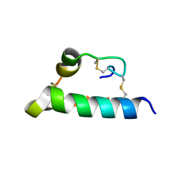
Solution NMR structure of the N-terminal immunoglobulin variable domain of BTNL2
Descriptor: Butyrophilin-like protein 2
Authors:Basak, A.J, Lee, W, Samanta, D, De, S.
Deposit date:2019-11-03
Release date:2020-10-14
Last modified:2023-06-14
Method:SOLUTION NMR
Cite:Structural Insights into N-terminal IgV Domain of BTNL2, a T Cell Inhibitory Molecule, Suggests a Non-canonical Binding Interface for Its Putative Receptors.
J.Mol.Biol., 432, 2020
6IGS
DownloadVisualize
BU of 6igs by Molmil
Crystal structure of HPRT from F. tularensis with Zinc
Descriptor: Hypoxanthine phosphoribosyltransferase, SULFATE ION, ZINC ION
Authors:Pavithra, G.C, Kundapura, S.V, Ramagopal, U.A.
Deposit date:2018-09-25
Release date:2019-10-02
Last modified:2023-11-22
Method:X-RAY DIFFRACTION (2.16 Å)
Cite:Crystal structure of hypoxanthine phosphoribosyltransferase from Francisella tularensis
To be published

220113

PDB entries from 2024-05-22

PDB statisticsPDBj update infoContact PDBjnumon