Loading
PDBj
MenuPDBj@FacebookPDBj@TwitterPDBj@YouTubewwPDB FoundationwwPDB
RCSB PDBPDBeBMRBAdv. SearchSearch help
Search by PDB author
5JOS
DownloadVisualize
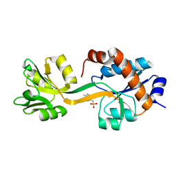
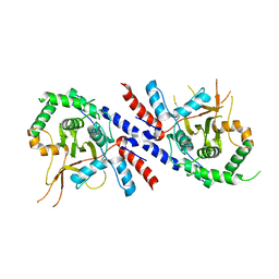
BU of 5jos by Molmil
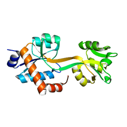
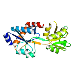
Crystal structure of an ancestral cyclohexadienyl dehydratase, AncCDT-3(P188L).
Descriptor: BENZOIC ACID, CITRIC ACID, Cyclohexadienyl dehydratase, ...
Authors:Clifton, B.E, Carr, P.D, Jackson, C.J.
Deposit date:2016-05-03
Release date:2017-05-03
Last modified:2023-09-27
Method:X-RAY DIFFRACTION (2.1 Å)
Cite:To be published
To Be Published
6BQE
DownloadVisualize
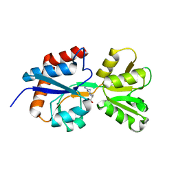
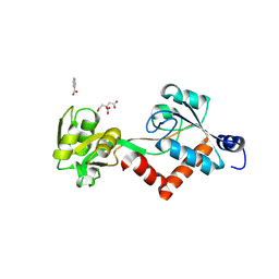
BU of 6bqe by Molmil
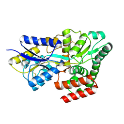
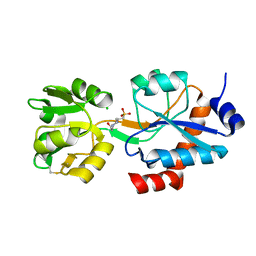
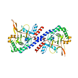
Low-resolution structure of cyclohexadienyl dehydratase from Pseudomonas aeruginosa in space group P4322.
Descriptor: ACETATE ION, Arogenate dehydratase
Authors:Clifton, B.E, Carr, P.D, Jackson, C.J.
Deposit date:2017-11-27
Release date:2017-12-13
Last modified:2018-05-30
Method:X-RAY DIFFRACTION (3.2 Å)
Cite:Evolution of cyclohexadienyl dehydratase from an ancestral solute-binding protein.
Nat. Chem. Biol., 14, 2018
5WJP
DownloadVisualize
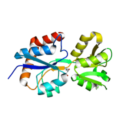
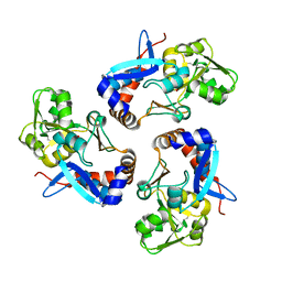
BU of 5wjp by Molmil
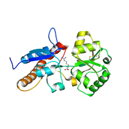
Crystal structure of the cyclohexadienyl dehydratase-like solute-binding protein SAR11_1068 from Candidatus Pelagibacter ubique.
Descriptor: Cyclohexadienyl dehydratase
Authors:Clifton, B.E, Jackson, C.J.
Deposit date:2017-07-24
Release date:2017-08-02
Last modified:2023-10-04
Method:X-RAY DIFFRACTION (1.57 Å)
Cite:Evolution of cyclohexadienyl dehydratase from an ancestral solute-binding protein.
Nat. Chem. Biol., 14, 2018
8HQQ
DownloadVisualize
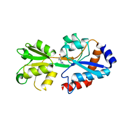
BU of 8hqq by Molmil
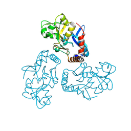
Crystal structure of the glucose-binding protein SAR11_0769 from "Candidatus Pelagibacter ubique" HTCC1062 bound to glucose
Descriptor: Probable binding protein component of ABC sugar transporter, beta-D-glucopyranose
Authors:Clifton, B.E, Laurino, P.
Deposit date:2022-12-14
Release date:2023-12-27
Method:X-RAY DIFFRACTION (1.86 Å)
Cite:Crystal structure of the glucose-binding protein SAR11_0769 from "Candidatus Pelagibacter ubique" HTCC1062 bound to glucose
To Be Published
8HQR
DownloadVisualize
BU of 8hqr by Molmil
Crystal structure of the arginine-/lysine-binding protein SAR11_1210 from 'Candidatus Pelagibacter ubique' HTCC1062 bound to arginine
Descriptor: ABC transporter, ARGININE
Authors:Clifton, B.E, Laurino, P.
Deposit date:2022-12-14
Release date:2023-12-27
Method:X-RAY DIFFRACTION (1.32 Å)
Cite:Crystal structure of the arginine-/lysine-binding protein SAR11_1210 from 'Candidatus Pelagibacter ubique' HTCC1062 bound to arginine
To Be Published
5HPQ
DownloadVisualize
BU of 5hpq by Molmil
Crystal structure of cyclohexadienyl dehydratase from Pseudomonas aeruginosa bound to acetate
Descriptor: ACETATE ION, Cyclohexadienyl dehydratase
Authors:Clifton, B.E, Carr, P.D, Jackson, C.J.
Deposit date:2016-01-20
Release date:2017-01-25
Last modified:2023-09-27
Method:X-RAY DIFFRACTION (2.05 Å)
Cite:Crystal structure of cyclohexadienyl dehydratase from Pseudomonas aeruginosa bound to acetate
To Be Published
5KKW
DownloadVisualize
BU of 5kkw by Molmil
Crystal structure of SAR11_1068 bound to a sulfobetaine (3-(1-methylpiperidinium-1-yl)propane-1-sulfonate)
Descriptor: 3-(1-methylpiperidinium-1-yl)propane-1-sulfonate, Cyclohexadienyl dehydratase, SULFATE ION
Authors:Clifton, B.E, Carr, P.D, Jackson, C.J.
Deposit date:2016-06-22
Release date:2017-07-05
Last modified:2023-09-27
Method:X-RAY DIFFRACTION (1.88 Å)
Cite:Crystal structure of SAR11_1068 bound to a sulfobetaine (3-(1-methylpiperidinium-1-yl)propane-1-sulfonate)
To Be Published
4ZV1
DownloadVisualize
BU of 4zv1 by Molmil
An ancestral arginine-binding protein bound to arginine
Descriptor: ARGININE, AncQR
Authors:Clifton, B.E, Carr, P.D, Jackson, C.J.
Deposit date:2015-05-18
Release date:2016-02-03
Last modified:2023-09-27
Method:X-RAY DIFFRACTION (1.52 Å)
Cite:Ancestral Protein Reconstruction Yields Insights into Adaptive Evolution of Binding Specificity in Solute-Binding Proteins.
Cell Chem Biol, 23, 2016
4ZV2
DownloadVisualize
BU of 4zv2 by Molmil
An ancestral arginine-binding protein bound to glutamine
Descriptor: AncQR, GLUTAMINE
Authors:Clifton, B.E, Jackson, C.J.
Deposit date:2015-05-18
Release date:2016-02-03
Last modified:2023-09-27
Method:X-RAY DIFFRACTION (1.43 Å)
Cite:Ancestral Protein Reconstruction Yields Insights into Adaptive Evolution of Binding Specificity in Solute-Binding Proteins.
Cell Chem Biol, 23, 2016
5T0W
DownloadVisualize
BU of 5t0w by Molmil
Crystal structure of the ancestral amino acid-binding protein AncCDT-1, a precursor of cyclohexadienyl dehydratase
Descriptor: ARGININE, AncCDT-1
Authors:Clifton, B.E, Carr, P.D, Jackson, C.J.
Deposit date:2016-08-16
Release date:2017-09-06
Last modified:2023-10-04
Method:X-RAY DIFFRACTION (2.59 Å)
Cite:To be published
To Be Published
5TUJ
DownloadVisualize
BU of 5tuj by Molmil
Ancestral Cationic Amino Acid Solute Binding Protein (AncCDT-1)
Descriptor: Ancestral protein CDT-Anc1
Authors:Kaczmarski, J.A, Clifton, B.E, Carr, P.D, Jackson, C.J.
Deposit date:2016-11-06
Release date:2017-12-06
Last modified:2024-03-06
Method:X-RAY DIFFRACTION (3.352 Å)
Cite:Evolution of cyclohexadienyl dehydratase from an ancestral solute-binding protein.
Nat. Chem. Biol., 14, 2018
6WUP
DownloadVisualize
BU of 6wup by Molmil
Crystal structure of an ancestral cyclohexadienyl dehydratase, AncCDT-5
Descriptor: 4-(2-HYDROXYETHYL)-1-PIPERAZINE ETHANESULFONIC ACID, Ancestral cyclohexadienyl dehydratase, AncCDT-5, ...
Authors:Kaczmarski, J.A, Mahawaththa, M.C.
Deposit date:2020-05-05
Release date:2020-05-13
Last modified:2023-10-18
Method:X-RAY DIFFRACTION (1.49 Å)
Cite:Altered conformational sampling along an evolutionary trajectory changes the catalytic activity of an enzyme.
Nat Commun, 11, 2020
7KWW
DownloadVisualize
BU of 7kww by Molmil
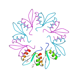
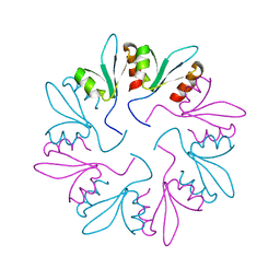
X-ray Crystal Structure of PlyCB Mutant K59H
Descriptor: (4S)-2-METHYL-2,4-PENTANEDIOL, PlyCB
Authors:Williams, D.E, Broendum, S.S, Hayes, B.K, Drinkwater, N, McGowan, S.
Deposit date:2020-12-02
Release date:2021-04-07
Last modified:2023-10-18
Method:X-RAY DIFFRACTION (1.8 Å)
Cite:High avidity drives the interaction between the streptococcal C1 phage endolysin, PlyC, with the cell surface carbohydrates of Group A Streptococcus.
Mol.Microbiol., 116, 2021
7KWY
DownloadVisualize
BU of 7kwy by Molmil
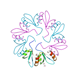
X-ray Crystal Structure of PlyCB Mutant R66K
Descriptor: (4S)-2-METHYL-2,4-PENTANEDIOL, PlyCB
Authors:Williams, D.E, Broendum, S.S, Hayes, B.K, Drinkwater, N, McGowan, S.
Deposit date:2020-12-02
Release date:2021-04-07
Last modified:2023-10-18
Method:X-RAY DIFFRACTION (1.7 Å)
Cite:High avidity drives the interaction between the streptococcal C1 phage endolysin, PlyC, with the cell surface carbohydrates of Group A Streptococcus.
Mol.Microbiol., 116, 2021
7KWT
DownloadVisualize
BU of 7kwt by Molmil
X-ray Crystal Structure of PlyCB Mutant Y28H
Descriptor: PlyCB
Authors:Williams, D.E, Broendum, S.S, Hayes, B.K, Drinkwater, N, McGowan, S.
Deposit date:2020-12-02
Release date:2021-04-07
Last modified:2023-10-18
Method:X-RAY DIFFRACTION (1.79 Å)
Cite:High avidity drives the interaction between the streptococcal C1 phage endolysin, PlyC, with the cell surface carbohydrates of Group A Streptococcus.
Mol.Microbiol., 116, 2021
5V3B
DownloadVisualize
BU of 5v3b by Molmil
Human A20 OTU domain (WT) with acetamidylated C103
Descriptor: Tumor necrosis factor alpha-induced protein 3
Authors:Langley, D.B, Christ, D, Grey, S.
Deposit date:2017-03-07
Release date:2018-03-21
Last modified:2023-10-04
Method:X-RAY DIFFRACTION (3 Å)
Cite:Denisovan, modern human and mouse TNFAIP3 alleles tune A20 phosphorylation and immunity.
Nat.Immunol., 20, 2019
4N1E
DownloadVisualize
BU of 4n1e by Molmil
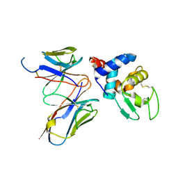
Structural evidence for antigen receptor evolution
Descriptor: Lysozyme C, immunoglobulin variable light chain domain
Authors:Langley, D.B, Rouet, R, Stock, D, Christ, D.
Deposit date:2013-10-04
Release date:2014-10-29
Last modified:2018-04-18
Method:X-RAY DIFFRACTION (2.23 Å)
Cite:Structural reconstruction of protein ancestry.
Proc. Natl. Acad. Sci. U.S.A., 114, 2017
4N1C
DownloadVisualize
BU of 4n1c by Molmil
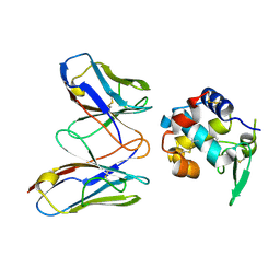
Structural evidence for antigen receptor evolution
Descriptor: Lysozyme C, immunoglobulin variable light chain domain
Authors:Langley, D.B, Rouet, R, Roome, B, Stock, D, Christ, D.
Deposit date:2013-10-03
Release date:2014-10-29
Last modified:2023-09-20
Method:X-RAY DIFFRACTION (1.7 Å)
Cite:Structural reconstruction of protein ancestry.
Proc. Natl. Acad. Sci. U.S.A., 114, 2017
5V3P
DownloadVisualize
BU of 5v3p by Molmil
Human A20 OTU domain (I325N) with acetamidylated C103
Descriptor: Tumor necrosis factor alpha-induced protein 3
Authors:Langley, D.B, Christ, D, Grey, S.
Deposit date:2017-03-07
Release date:2018-03-21
Last modified:2023-10-04
Method:X-RAY DIFFRACTION (2.5 Å)
Cite:Denisovan, modern human and mouse TNFAIP3 alleles tune A20 phosphorylation and immunity.
Nat.Immunol., 20, 2019

219140

PDB entries from 2024-05-01

PDB statisticsPDBj update infoContact PDBjnumon