ムービー
ムービー コントローラー


+ データを開く

- 基本情報
基本情報
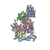
| 登録情報 | データベース: PDB / ID: 8ru2 | ||||||||||||
|---|---|---|---|---|---|---|---|---|---|---|---|---|---|
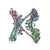
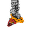
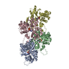
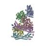
| タイトル | Structure of the F-actin barbed end bound by formin mDia1 | ||||||||||||
 要素 要素 |
| ||||||||||||
 キーワード キーワード |  STRUCTURAL PROTEIN (タンパク質) / actin (アクチン) / formin / Cdc12 / profilin (プロフィリン) / actin assembly (アクチン) STRUCTURAL PROTEIN (タンパク質) / actin (アクチン) / formin / Cdc12 / profilin (プロフィリン) / actin assembly (アクチン) | ||||||||||||
| 機能・相同性 |  機能・相同性情報 機能・相同性情報negative regulation of neuron projection regeneration / multicellular organismal locomotion / MGMT-mediated DNA damage reversal / RHOF GTPase cycle / RHOB GTPase cycle / ERBB2 Regulates Cell Motility / RHOC GTPase cycle / RHOD GTPase cycle / RHOA GTPase cycle / methylated-DNA-[protein]-cysteine S-methyltransferase ...negative regulation of neuron projection regeneration / multicellular organismal locomotion / MGMT-mediated DNA damage reversal / RHOF GTPase cycle / RHOB GTPase cycle / ERBB2 Regulates Cell Motility / RHOC GTPase cycle / RHOD GTPase cycle / RHOA GTPase cycle / methylated-DNA-[protein]-cysteine S-methyltransferase / methylated-DNA-[protein]-cysteine S-methyltransferase activity / actin nucleation / positive regulation of norepinephrine uptake / neuron projection retraction / cellular response to cytochalasin B / DNA-methyltransferase activity / bBAF complex / npBAF complex / RHO GTPases Activate Formins / protein localization to microtubule / postsynaptic actin cytoskeleton organization / regulation of transepithelial transport / brahma complex / nBAF complex / structural constituent of postsynaptic actin cytoskeleton / profilin binding / morphogenesis of a polarized epithelium / cellular response to histamine / GBAF complex / postsynaptic actin cytoskeleton / protein localization to adherens junction / Formation of annular gap junctions / regulation of G0 to G1 transition / dense body / Gap junction degradation / Tat protein binding / DNA alkylation repair / Cell-extracellular matrix interactions / Folding of actin by CCT/TriC / regulation of double-strand break repair / DNA ligation / regulation of nucleotide-excision repair / RSC-type complex / apical protein localization / Prefoldin mediated transfer of substrate to CCT/TriC / regulation of microtubule-based process / regulation of release of sequestered calcium ion into cytosol / adherens junction assembly / RHOF GTPase cycle / Adherens junctions interactions / axon midline choice point recognition / 密着結合 / Sensory processing of sound by outer hair cells of the cochlea / SWI/SNF complex / Interaction between L1 and Ankyrins / Sensory processing of sound by inner hair cells of the cochlea / regulation of mitotic metaphase/anaphase transition / regulation of norepinephrine uptake / positive regulation of double-strand break repair / positive regulation of T cell differentiation / NuA4 histone acetyltransferase complex / regulation of synaptic vesicle endocytosis / regulation of cytoskeleton organization / apical junction complex / establishment or maintenance of cell polarity / maintenance of blood-brain barrier / positive regulation of stem cell population maintenance / positive regulation of double-strand break repair via homologous recombination / cortical cytoskeleton / nitric-oxide synthase binding / regulation of cyclin-dependent protein serine/threonine kinase activity / Recycling pathway of L1 / regulation of G1/S transition of mitotic cell cycle / negative regulation of cell differentiation / 刷子縁 / kinesin binding / ヘルト萼状シナプス / synaptic vesicle endocytosis / EPH-ephrin mediated repulsion of cells / RHO GTPases Activate WASPs and WAVEs / ephrin receptor signaling pathway / RHO GTPases activate IQGAPs / positive regulation of myoblast differentiation / regulation of protein localization to plasma membrane / cytoskeleton organization / EPHB-mediated forward signaling / substantia nigra development / actin filament polymerization / Neutrophil degranulation / 軸索誘導 / negative regulation of protein binding / 運動性 / methyltransferase activity / マイクロフィラメント / Translocation of SLC2A4 (GLUT4) to the plasma membrane / RHO GTPases Activate Formins / regulation of transmembrane transporter activity / positive regulation of cell differentiation / sensory perception of sound / FCGR3A-mediated phagocytosis類似検索 - 分子機能 | ||||||||||||
| 生物種 |  Homo sapiens (ヒト) Homo sapiens (ヒト) Mus musculus (ハツカネズミ) Mus musculus (ハツカネズミ) | ||||||||||||
| 手法 | 電子顕微鏡法 / 単粒子再構成法 / クライオ電子顕微鏡法 / 解像度: 3.49 Å | ||||||||||||
 データ登録者 データ登録者 | Oosterheert, W. / Boiero Sanders, M. / Funk, J. / Prumbaum, D. / Raunser, S. / Bieling, P. | ||||||||||||
| 資金援助 |  ドイツ, European Union, 3件 ドイツ, European Union, 3件
| ||||||||||||
 引用 引用 | ジャーナル: Science / 年: 2024 タイトル: Molecular mechanism of actin filament elongation by formins. 著者: Wout Oosterheert / Micaela Boiero Sanders / Johanna Funk / Daniel Prumbaum / Stefan Raunser / Peter Bieling / 要旨: Formins control the assembly of actin filaments (F-actin) that drive cell morphogenesis and motility in eukaryotes. However, their molecular interaction with F-actin and their mechanism of action ...Formins control the assembly of actin filaments (F-actin) that drive cell morphogenesis and motility in eukaryotes. However, their molecular interaction with F-actin and their mechanism of action remain unclear. In this work, we present high-resolution cryo-electron microscopy structures of F-actin barbed ends bound by three distinct formins, revealing a common asymmetric formin conformation imposed by the filament. Formation of new intersubunit contacts during actin polymerization sterically displaces formin and triggers its translocation. This "undock-and-lock" mechanism explains how actin-filament growth is coordinated with formin movement. Filament elongation speeds are controlled by the positioning and stability of actin-formin interfaces, which distinguish fast and slow formins. Furthermore, we provide a structure of the actin-formin-profilin ring complex, which resolves how profilin is rapidly released from the barbed end during filament elongation. | ||||||||||||
| 履歴 |
|
- 構造の表示
構造の表示
| 構造ビューア | 分子: MolmilJmol/JSmol |
|---|
- ダウンロードとリンク
-ダウンロード
| PDBx/mmCIF形式 | 8ru2.cif.gz | 346.2 KB | 表示 | PDBx/mmCIF形式 |
|---|---|---|---|---|
| PDB形式 | pdb8ru2.ent.gz | 271.8 KB | 表示 | PDB形式 |
| PDBx/mmJSON形式 | 8ru2.json.gz | ツリー表示 | PDBx/mmJSON形式 | |
| その他 |  その他のダウンロード その他のダウンロード |
-検証レポート
| アーカイブディレクトリ | https://data.pdbj.org/pub/pdb/validation_reports/ru/8ru2ftp://data.pdbj.org/pub/pdb/validation_reports/ru/8ru2 | HTTPS FTP |
|---|
-関連構造データ


-リンク
 PDBj
PDBj


- 集合体
集合体
| 登録構造単位 | 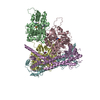
|
|---|---|
| 1 |
|
-要素
| #1: タンパク質 | アクチン 分子量: 41632.422 Da / 分子数: 3 / 由来タイプ: 組換発現 詳細: Human beta-actin was recombinantly purified from BTI-Tnao38 cells. 由来: (組換発現) Homo sapiens (ヒト) / 遺伝子: ACTB / プラスミド: p2336 pFL_ACTB_C272A / 細胞株 (発現宿主): BTI-Tnao38 / 発現宿主:  Trichoplusia ni (イラクサキンウワバ) / 参照: UniProt: P60709 Trichoplusia ni (イラクサキンウワバ) / 参照: UniProt: P60709#2: タンパク質 | 分子量: 86469.258 Da / 分子数: 2 / 由来タイプ: 組換発現 / 詳細: Has a N-terminal snap-tag. 由来: (組換発現) Homo sapiens (ヒト), (組換発現) Mus musculus (ハツカネズミ)遺伝子: MGMT, Diaph1, Diap1 / 発現宿主:  Escherichia coli (大腸菌) / 株 (発現宿主): BL21 Star pRARE Escherichia coli (大腸菌) / 株 (発現宿主): BL21 Star pRARE参照: UniProt: P16455, UniProt: O08808, methylated-DNA-[protein]-cysteine S-methyltransferase #3: 化合物 | アデノシン二リン酸  分子量: 427.201 Da / 分子数: 3 / 由来タイプ: 合成 / 式: C10H15N5O10P2 / タイプ: SUBJECT OF INVESTIGATION / コメント: ADP, エネルギー貯蔵分子*YM 分子量: 427.201 Da / 分子数: 3 / 由来タイプ: 合成 / 式: C10H15N5O10P2 / タイプ: SUBJECT OF INVESTIGATION / コメント: ADP, エネルギー貯蔵分子*YM#4: 化合物 |   分子量: 24.305 Da / 分子数: 3 / 由来タイプ: 合成 / 式: Mg 分子量: 24.305 Da / 分子数: 3 / 由来タイプ: 合成 / 式: Mg研究の焦点であるリガンドがあるか | Y | |
|---|
-実験情報
-実験
| 実験 | 手法: 電子顕微鏡法 |
|---|---|
| EM実験 | 試料の集合状態: PARTICLE / 3次元再構成法: 単粒子再構成法 |
- 試料調製
試料調製
| 構成要素 |
| ||||||||||||||||||||||||||||
|---|---|---|---|---|---|---|---|---|---|---|---|---|---|---|---|---|---|---|---|---|---|---|---|---|---|---|---|---|---|
| 分子量 | 実験値: NO | ||||||||||||||||||||||||||||
| 由来(天然) |
| ||||||||||||||||||||||||||||
| 由来(組換発現) |
| ||||||||||||||||||||||||||||
| 緩衝液 | pH: 7.1 | ||||||||||||||||||||||||||||
| 緩衝液成分 |
| ||||||||||||||||||||||||||||
| 試料 | 包埋: NO / シャドウイング: NO / 染色: NO / 凍結: YES | ||||||||||||||||||||||||||||
| 試料支持 | グリッドの材料: GOLD / グリッドのサイズ: 200 divisions/in. / グリッドのタイプ: Quantifoil R2/1 | ||||||||||||||||||||||||||||
| 急速凍結 | 装置: FEI VITROBOT MARK IV / 凍結剤: ETHANE-PROPANE / 湿度: 100 % / 凍結前の試料温度: 286 K / 詳細: 3 seconds, force 0. |
- 電子顕微鏡撮影
| 実験機器 |  モデル: Titan Krios / 画像提供: FEI Company |
|---|---|
| 顕微鏡 | モデル: FEI TITAN KRIOS 詳細: 300 kV Titan Krios G2 microscope (Thermo Fisher Scientific) with an in-column Cs-corrector. |
| 電子銃 | 電子線源: FIELD EMISSION GUN / 加速電圧: 300 kV / 照射モード: FLOOD BEAM |
| 電子レンズ | モード: BRIGHT FIELDBright-field microscopy / 倍率(公称値): 81000 X / 最大 デフォーカス(公称値): 2700 nm / 最小 デフォーカス(公称値): 1200 nm / Cs: 0.01 mm / C2レンズ絞り径: 50 µm |
| 試料ホルダ | 凍結剤: NITROGEN 試料ホルダーモデル: FEI TITAN KRIOS AUTOGRID HOLDER |
| 撮影 | 電子線照射量: 67.6 e/Å2 フィルム・検出器のモデル: GATAN K3 BIOQUANTUM (6k x 4k) 撮影したグリッド数: 2 / 実像数: 38913 |
| 電子光学装置 | エネルギーフィルター名称: GIF Bioquantum / 詳細: Gatan energy filter. / エネルギーフィルタースリット幅: 15 eV 球面収差補正装置 : Titan Krios G2 microscope (Thermo Fisher Scientific) with an in-column Cs-corrector. |
- 解析
解析
| EMソフトウェア |
| ||||||||||||||||||||||||||||||||||||||||||||||||
|---|---|---|---|---|---|---|---|---|---|---|---|---|---|---|---|---|---|---|---|---|---|---|---|---|---|---|---|---|---|---|---|---|---|---|---|---|---|---|---|---|---|---|---|---|---|---|---|---|---|
| CTF補正 | タイプ: PHASE FLIPPING AND AMPLITUDE CORRECTION | ||||||||||||||||||||||||||||||||||||||||||||||||
| 粒子像の選択 | 選択した粒子像数: 1963686 / 詳細: Particles picked using SPHIRE-crYOLO. | ||||||||||||||||||||||||||||||||||||||||||||||||
| 対称性 | 点対称性: C1 (非対称) | ||||||||||||||||||||||||||||||||||||||||||||||||
| 3次元再構成 | 解像度: 3.49 Å / 解像度の算出法: FSC 0.143 CUT-OFF / 粒子像の数: 150807 / 対称性のタイプ: POINT | ||||||||||||||||||||||||||||||||||||||||||||||||
| 原子モデル構築 | プロトコル: FLEXIBLE FIT / 空間: REAL / 詳細: Refinement performed using phenix real-space refine | ||||||||||||||||||||||||||||||||||||||||||||||||
| 原子モデル構築 | 3D fitting-ID: 1 / Source name: PDB / タイプ: experimental model
| ||||||||||||||||||||||||||||||||||||||||||||||||
| 精密化 | 交差検証法: NONE 立体化学のターゲット値: GeoStd + Monomer Library + CDL v1.2 | ||||||||||||||||||||||||||||||||||||||||||||||||
| 原子変位パラメータ | Biso mean: 115.26 Å2 | ||||||||||||||||||||||||||||||||||||||||||||||||
| 拘束条件 |
|