 Movie
Movie Controller


+ Open data

- Basic information
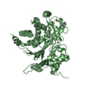
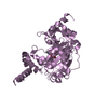
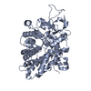
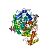
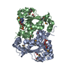
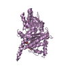
| Entry | Database: PDB / ID: 7c7r | ||||||
|---|---|---|---|---|---|---|---|
| Title | Biofilm associated protein - B domain | ||||||
 Components Components | Biofilm-associated surface protein | ||||||
 Keywords Keywords |  CELL ADHESION / Cell wall-anchored surface protein / biofilm formation CELL ADHESION / Cell wall-anchored surface protein / biofilm formation | ||||||
| Function / homology |  Function and homology information Function and homology informationcell-abiotic substrate adhesion / single-species submerged biofilm formation / cell-cell adhesion / membraneSimilarity search - Function | ||||||
| Biological species |   Staphylococcus aureus (bacteria) Staphylococcus aureus (bacteria) | ||||||
| Method | X-RAY DIFFRACTION / SYNCHROTRON / SAD / Resolution: 3.07 Å | ||||||
 Authors Authors | Ma, J.F. / Xu, Z.H. / Zhang, Y.K. / Cheng, X. / Fan, S.L. / Wang, J.W. / Fang, X.Y. | ||||||
| Funding support |  China, 1items China, 1items
| ||||||
 Citation Citation | Journal: Embo J. / Year: 2021 Title: Structural mechanism for modulation of functional amyloid and biofilm formation by Staphylococcal Bap protein switch. Authors: Ma, J. / Cheng, X. / Xu, Z. / Zhang, Y. / Valle, J. / Fan, S. / Zuo, X. / Lasa, I. / Fang, X. #1: Journal: To Be PublishedTitle: Integrative structure and functional implications of a 236-kDa modular protein Bap from Staphylococcus aureus Authors: Ma, J.F. / Zhang, Y.K. / Cheng, X. / Fan, S.L. / Xu, N. / Fang, X.Y. | ||||||
| History |
|
- Structure visualization
| Structure viewer | Molecule: MolmilJmol/JSmol |
|---|
- Downloads & links
-Download
| PDBx/mmCIF format | 7c7r.cif.gz | 336.3 KB | Display | PDBx/mmCIF format |
|---|---|---|---|---|
| PDB format | pdb7c7r.ent.gz | 283.8 KB | Display | PDB format |
| PDBx/mmJSON format | 7c7r.json.gz | Tree view | PDBx/mmJSON format | |
| Others |  Other downloads Other downloads |
-Validation report
| Arichive directory | https://data.pdbj.org/pub/pdb/validation_reports/c7/7c7rftp://data.pdbj.org/pub/pdb/validation_reports/c7/7c7r | HTTPS FTP |
|---|
-Related structure data


-Links
 PDBj
PDBj- Assembly
| Deposited unit | 
| |||||||||||||||||||||||||||||||||||||||||||||||||||
|---|---|---|---|---|---|---|---|---|---|---|---|---|---|---|---|---|---|---|---|---|---|---|---|---|---|---|---|---|---|---|---|---|---|---|---|---|---|---|---|---|---|---|---|---|---|---|---|---|---|---|---|---|
| 1 | 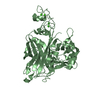
| |||||||||||||||||||||||||||||||||||||||||||||||||||
| 2 | 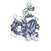
| |||||||||||||||||||||||||||||||||||||||||||||||||||
| Unit cell |
| |||||||||||||||||||||||||||||||||||||||||||||||||||
| Noncrystallographic symmetry (NCS) | NCS domain:
NCS domain segments:
|
-Components
| #1: Protein | Mass: 49767.359 Da / Num. of mol.: 2 Source method: isolated from a genetically manipulated source Source: (gene. exp.) Staphylococcus aureus (bacteria) / Gene: bap / Production host: Escherichia coli BL21(DE3) (bacteria) / Strain (production host): BL21(DE3) / Variant (production host): B834 / References: UniProt: Q79LN3#2: Chemical | ChemComp-CA /   Mass: 40.078 Da / Num. of mol.: 10 / Source method: obtained synthetically / Formula: Ca / Feature type: SUBJECT OF INVESTIGATION Mass: 40.078 Da / Num. of mol.: 10 / Source method: obtained synthetically / Formula: Ca / Feature type: SUBJECT OF INVESTIGATION#3: Water | ChemComp-HOH / | Water Mass: 18.015 Da / Num. of mol.: 30 / Source method: isolated from a natural source / Formula: H2O Mass: 18.015 Da / Num. of mol.: 30 / Source method: isolated from a natural source / Formula: H2OHas ligand of interest | Y | |
|---|
-Experimental details
-Experiment
| Experiment | Method: X-RAY DIFFRACTION / Number of used crystals: 1 |
|---|
- Sample preparation
| Crystal | Density Matthews: 4.06 Å3/Da / Density % sol: 69.67 % |
|---|---|
| Crystal grow | Temperature: 289.15 K / Method: vapor diffusion, hanging drop / Details: 0.1M Sodium Cacodylate pH 6.5, 40% MPD, 5% PEG8000 |
-Data collection
| Diffraction | Mean temperature: 100 K / Serial crystal experiment: N |
|---|---|
| Diffraction source | Source: SYNCHROTRON / Site: SSRF / Beamline: BL19U1 / Wavelength: 0.97892 Å |
| Detector | Type: DECTRIS PILATUS 6M / Detector: PIXEL / Date: Jul 11, 2018 |
| Radiation | Protocol: SINGLE WAVELENGTH / Monochromatic (M) / Laue (L): M / Scattering type: x-ray |
| Radiation wavelength | Wavelength: 0.97892 Å / Relative weight: 1 |
| Reflection | Resolution: 3.07→49.1 Å / Num. obs: 27970 / % possible obs: 96.42 % / Redundancy: 2 % / Biso Wilson estimate: 76.02 Å2 / CC1/2: 0.999 / Net I/σ(I): 25.33 |
| Reflection shell | Resolution: 3.07→3.18 Å / Num. unique obs: 2317 / CC1/2: 0.661 |
- Processing
| Software |
| |||||||||||||||||||||||||||||||||||||||||||||||||||||||||||||||||||||||||||||
|---|---|---|---|---|---|---|---|---|---|---|---|---|---|---|---|---|---|---|---|---|---|---|---|---|---|---|---|---|---|---|---|---|---|---|---|---|---|---|---|---|---|---|---|---|---|---|---|---|---|---|---|---|---|---|---|---|---|---|---|---|---|---|---|---|---|---|---|---|---|---|---|---|---|---|---|---|---|---|
| Refinement | Method to determine structure: SAD / Resolution: 3.07→49.1 Å / SU ML: 0.36 / Cross valid method: THROUGHOUT / σ(F): 1.34 / Phase error: 22.89 / Stereochemistry target values: ML
| |||||||||||||||||||||||||||||||||||||||||||||||||||||||||||||||||||||||||||||
| Solvent computation | Shrinkage radii: 0.9 Å / VDW probe radii: 1.11 Å / Solvent model: FLAT BULK SOLVENT MODEL | |||||||||||||||||||||||||||||||||||||||||||||||||||||||||||||||||||||||||||||
| Displacement parameters | Biso max: 168.71 Å2 / Biso mean: 71.6052 Å2 / Biso min: 25.82 Å2 | |||||||||||||||||||||||||||||||||||||||||||||||||||||||||||||||||||||||||||||
| Refinement step | Cycle: final / Resolution: 3.07→49.1 Å
| |||||||||||||||||||||||||||||||||||||||||||||||||||||||||||||||||||||||||||||
| Refine LS restraints NCS |
| |||||||||||||||||||||||||||||||||||||||||||||||||||||||||||||||||||||||||||||
| LS refinement shell | Refine-ID: X-RAY DIFFRACTION / Rfactor Rfree error: 0 / Total num. of bins used: 10
| |||||||||||||||||||||||||||||||||||||||||||||||||||||||||||||||||||||||||||||
| Refinement TLS params. | Method: refined / Origin x: 139.4015 Å / Origin y: 62.7425 Å / Origin z: 24.636 Å
| |||||||||||||||||||||||||||||||||||||||||||||||||||||||||||||||||||||||||||||
| Refinement TLS group |
|
