 Movie
Movie Controller

[English] 日本語
 Yorodumi
- PDB-2rjb: Crystal structure of uncharacterized protein YdcJ (SF1787) from S... -
+ Open data

- Basic information
| Entry | Database: PDB / ID: 2rjb | ||||||
|---|---|---|---|---|---|---|---|
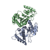
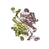
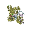
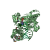
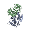
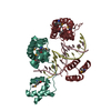
| Title | Crystal structure of uncharacterized protein YdcJ (SF1787) from Shigella flexneri which includes domain DUF1338. Northeast Structural Genomics Consortium target SfR276 | ||||||
 Components Components | Uncharacterized protein | ||||||
 Keywords Keywords |  STRUCTURAL GENOMICS / UNKNOWN FUNCTION / Uncharacterized protein / PSI-2 / Protein Structure Initiative / Northeast Structural Genomics Consortium / NESG STRUCTURAL GENOMICS / UNKNOWN FUNCTION / Uncharacterized protein / PSI-2 / Protein Structure Initiative / Northeast Structural Genomics Consortium / NESG | ||||||
| Function / homology |  Function and homology information Function and homology information | ||||||
| Biological species |  Shigella flexneri (bacteria) Shigella flexneri (bacteria) | ||||||
| Method | X-RAY DIFFRACTION / SYNCHROTRON / SAD / Resolution: 2.6 Å | ||||||
 Authors Authors | Seetharaman, J. / Chen, Y. / Wang, D. / Fang, Y. / Cunningham, K. / Ma, L.-C. / Xia, R. / Liu, J. / Baran, M.C. / Acton, T.B. ...Seetharaman, J. / Chen, Y. / Wang, D. / Fang, Y. / Cunningham, K. / Ma, L.-C. / Xia, R. / Liu, J. / Baran, M.C. / Acton, T.B. / Rost, B. / Montelione, G.T. / Tong, L. / Hunt, J.F. / Northeast Structural Genomics Consortium (NESG) | ||||||
 Citation Citation | Journal: To be Published Title: Crystal structure of uncharacterized protein YdcJ (SF1787) from Shigella flexneri which includes domain DUF1338. Authors: Seetharaman, J. / Chen, Y. / Wang, D. / Fang, Y. / Cunningham, K. / Ma, L.-C. / Xia, R. / Liu, J. / Baran, M.C. / Acton, T.B. / Rost, B. / Montelione, G.T. / Tong, L. / Hunt, J.F. | ||||||
| History |
|
- Structure visualization
| Structure viewer | Molecule: MolmilJmol/JSmol |
|---|
- Downloads & links
-Download
| PDBx/mmCIF format | 2rjb.cif.gz | 327.4 KB | Display | PDBx/mmCIF format |
|---|---|---|---|---|
| PDB format | pdb2rjb.ent.gz | 277.7 KB | Display | PDB format |
| PDBx/mmJSON format | 2rjb.json.gz | Tree view | PDBx/mmJSON format | |
| Others |  Other downloads Other downloads |
-Validation report
| Arichive directory | https://data.pdbj.org/pub/pdb/validation_reports/rj/2rjbftp://data.pdbj.org/pub/pdb/validation_reports/rj/2rjb | HTTPS FTP |
|---|
-Related structure data
| Similar structure data | |
|---|---|
| Other databases |


-Links
 PDBj
PDBj- Assembly
| Deposited unit | 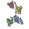
| ||||||||
|---|---|---|---|---|---|---|---|---|---|
| 1 | 
| ||||||||
| 2 | 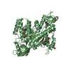
| ||||||||
| 3 | 
| ||||||||
| 4 | 
| ||||||||
| Unit cell |
|
-Components
| #1: Protein | Mass: 52714.539 Da / Num. of mol.: 4 Source method: isolated from a genetically manipulated source Source: (gene. exp.) Shigella flexneri (bacteria) / Strain: 301 / Serotype 2a / Gene: S1487, SF1787 / Plasmid: pET21 / Species (production host): Escherichia coli / Production host: Escherichia coli BL21(DE3) (bacteria) / Strain (production host): BL21(DE3) / References: UniProt: Q83KU0, UniProt: A0A0H2UZX2*PLUS#2: Chemical | ChemComp-ZN /   Mass: 65.409 Da / Num. of mol.: 4 / Source method: obtained synthetically / Formula: Zn Mass: 65.409 Da / Num. of mol.: 4 / Source method: obtained synthetically / Formula: Zn#3: Water | ChemComp-HOH / | Water Mass: 18.015 Da / Num. of mol.: 502 / Source method: isolated from a natural source / Formula: H2O Mass: 18.015 Da / Num. of mol.: 502 / Source method: isolated from a natural source / Formula: H2O |
|---|
-Experimental details
-Experiment
| Experiment | Method: X-RAY DIFFRACTION / Number of used crystals: 1 |
|---|
- Sample preparation
| Crystal | Density Matthews: 2.48 Å3/Da / Density % sol: 50.34 % Description: The structure factor file contains Friedel pairs |
|---|---|
| Crystal grow | Temperature: 277 K / Method: microbatch under oil / pH: 7 Details: NH4H2PO4, 0.1M MOPS, 20% PEG 1000, pH 7.0, MICROBATCH UNDER OIL, temperature 277K |
-Data collection
| Diffraction | Mean temperature: 100 K |
|---|---|
| Diffraction source | Source: SYNCHROTRON / Site: NSLS  / Beamline: X4C / Wavelength: 0.979 Å / Beamline: X4C / Wavelength: 0.979 Å |
| Detector | Type: MAR scanner 345 mm plate / Detector: IMAGE PLATE / Date: Aug 10, 2007 |
| Radiation | Protocol: SINGLE WAVELENGTH / Monochromatic (M) / Laue (L): M / Scattering type: x-ray |
| Radiation wavelength | Wavelength: 0.979 Å / Relative weight: 1 |
| Reflection | Resolution: 2.6→50 Å / Num. all: 64525 / Num. obs: 64525 / % possible obs: 98.9 % / Observed criterion σ(F): 0 / Observed criterion σ(I): 0 / Redundancy: 3.9 % / Biso Wilson estimate: 31.2 Å2 / Rmerge(I) obs: 0.072 / Rsym value: 0.066 / Net I/σ(I): 11.3 |
| Reflection shell | Resolution: 2.6→2.69 Å / Redundancy: 3.9 % / Rmerge(I) obs: 0.228 / Mean I/σ(I) obs: 10 / Num. unique all: 12337 / Rsym value: 0.275 / % possible all: 98.5 |
- Processing
| Software |
| ||||||||||||||||||||||||||||||||||||
|---|---|---|---|---|---|---|---|---|---|---|---|---|---|---|---|---|---|---|---|---|---|---|---|---|---|---|---|---|---|---|---|---|---|---|---|---|---|
| Refinement | Method to determine structure: SAD / Resolution: 2.6→41.12 Å / Rfactor Rfree error: 0.005 / Data cutoff high absF: 108028.4 / Data cutoff low absF: 0 / Isotropic thermal model: RESTRAINED / Cross valid method: THROUGHOUT / σ(F): 2 / Stereochemistry target values: Engh & Huber / Details: The Friedel pairs were used in phasing
| ||||||||||||||||||||||||||||||||||||
| Solvent computation | Solvent model: FLAT MODEL / Bsol: 27.9052 Å2 / ksol: 0.313935 e/Å3 | ||||||||||||||||||||||||||||||||||||
| Displacement parameters | Biso mean: 45.5 Å2
| ||||||||||||||||||||||||||||||||||||
| Refine analyze |
| ||||||||||||||||||||||||||||||||||||
| Refinement step | Cycle: LAST / Resolution: 2.6→41.12 Å
| ||||||||||||||||||||||||||||||||||||
| Refine LS restraints |
| ||||||||||||||||||||||||||||||||||||
| LS refinement shell | Resolution: 2.6→2.76 Å / Rfactor Rfree error: 0.017 / Total num. of bins used: 6
| ||||||||||||||||||||||||||||||||||||
| Xplor file |
|
