 Movie
Movie Controller

[English] 日本語
 Yorodumi
- PDB-5jjt: Crystal structure of a type 5 serine/threonine protein phosphatas... -
+ Open data

- Basic information
| Entry | Database: PDB / ID: 5jjt | ||||||
|---|---|---|---|---|---|---|---|
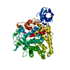
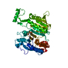
| Title | Crystal structure of a type 5 serine/threonine protein phosphatase from Arabidopsis thaliana | ||||||
 Components Components | Serine/threonine-protein phosphatase 5 | ||||||
 Keywords Keywords |  HYDROLASE / phosphatase HYDROLASE / phosphatase | ||||||
| Function / homology |  Function and homology information Function and homology informationnegative regulation of chlorophyll biosynthetic process / red or far-red light signaling pathway / chloroplast-nucleus signaling pathway / tetrapyrrole binding / plasmodesma / nucleocytoplasmic transport / myosin phosphatase activity / protein serine/threonine phosphatase activity / protein-serine/threonine phosphatase / phosphoprotein phosphatase activity ...negative regulation of chlorophyll biosynthetic process / red or far-red light signaling pathway / chloroplast-nucleus signaling pathway / tetrapyrrole binding / plasmodesma / nucleocytoplasmic transport / myosin phosphatase activity / protein serine/threonine phosphatase activity / protein-serine/threonine phosphatase / phosphoprotein phosphatase activity / nuclear envelope / nuclear membrane / nuclear speck / endoplasmic reticulum membrane / metal ion binding / nucleus / cytosol / cytoplasmSimilarity search - Function | ||||||
| Biological species |  Arabidopsis thaliana (thale cress) Arabidopsis thaliana (thale cress) | ||||||
| Method | X-RAY DIFFRACTION / SYNCHROTRON / MOLECULAR REPLACEMENT / Resolution: 2.103 Å | ||||||
 Authors Authors | Li, H.M. / Pu, H. | ||||||
 Citation Citation | Journal: To Be Published Title: Crystal structure of a type 5 serine/threonine protein phosphatase from Arabidopsis thaliana Authors: Li, H.M. / Pu, H. | ||||||
| History |
|
- Structure visualization
| Structure viewer | Molecule: MolmilJmol/JSmol |
|---|
- Downloads & links
-Download
| PDBx/mmCIF format | 5jjt.cif.gz | 216.3 KB | Display | PDBx/mmCIF format |
|---|---|---|---|---|
| PDB format | pdb5jjt.ent.gz | 168.9 KB | Display | PDB format |
| PDBx/mmJSON format | 5jjt.json.gz | Tree view | PDBx/mmJSON format | |
| Others |  Other downloads Other downloads |
-Validation report
| Arichive directory | https://data.pdbj.org/pub/pdb/validation_reports/jj/5jjtftp://data.pdbj.org/pub/pdb/validation_reports/jj/5jjt | HTTPS FTP |
|---|
-Related structure data
| Related structure data |  1waoS S: Starting model for refinement |
|---|---|
| Similar structure data |


-Links
 PDBj
PDBj


- Assembly
| Deposited unit | 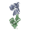
| ||||||||
|---|---|---|---|---|---|---|---|---|---|
| 1 | 
| ||||||||
| 2 | 
| ||||||||
| Unit cell |
|
-Components
| #1: Protein | Mass: 54169.391 Da / Num. of mol.: 2 / Fragment: UNP residues 5-483 of isoform 2 Source method: isolated from a genetically manipulated source Source: (gene. exp.) Arabidopsis thaliana (thale cress) / Gene: PAPP5, PP5, At2g42810, F7D19.19 / Production host:  Escherichia coli (E. coli) Escherichia coli (E. coli)References: UniProt: Q84XU2, protein-serine/threonine phosphatase #2: Chemical | ChemComp-NI / Nickel  Mass: 58.693 Da / Num. of mol.: 6 / Source method: isolated from a natural source / Formula: Ni Mass: 58.693 Da / Num. of mol.: 6 / Source method: isolated from a natural source / Formula: Ni#3: Water | ChemComp-HOH / | Water Mass: 18.015 Da / Num. of mol.: 770 / Source method: isolated from a natural source / Formula: H2O Mass: 18.015 Da / Num. of mol.: 770 / Source method: isolated from a natural source / Formula: H2O |
|---|
-Experimental details
-Experiment
| Experiment | Method: X-RAY DIFFRACTION / Number of used crystals: 1 |
|---|
- Sample preparation
| Crystal | Density Matthews: 2.31 Å3/Da / Density % sol: 46.8 % |
|---|---|
| Crystal grow | Temperature: 289 K / Method: vapor diffusion, sitting drop / pH: 7.2 / Details: 0.1M HEPES pH7.2, 22.5% PEG 3350, 20% glycol |
-Data collection
| Diffraction | Mean temperature: 100 K |
|---|---|
| Diffraction source | Source: SYNCHROTRON / Site: SSRF  / Beamline: BL17U / Wavelength: 0.9793 Å / Beamline: BL17U / Wavelength: 0.9793 Å |
| Detector | Type: ADSC QUANTUM 315r / Detector: CCD / Date: Jun 20, 2014 |
| Radiation | Protocol: SINGLE WAVELENGTH / Monochromatic (M) / Laue (L): M / Scattering type: x-ray |
| Radiation wavelength | Wavelength: 0.9793 Å / Relative weight: 1 |
| Reflection | Resolution: 2.1→50 Å / Num. obs: 55086 / % possible obs: 96.6 % / Redundancy: 3.5 % / Rmerge(I) obs: 0.091 / Net I/σ(I): 12.97 |
| Reflection shell | Resolution: 2.1→2.18 Å |
- Processing
| Software |
| |||||||||||||||||||||||||||||||||||||||||||||||||||||||||||||||||||||||||||||||||||||||||||||||||||||||||||||||||||||||||||||||||||||||||||||||||||
|---|---|---|---|---|---|---|---|---|---|---|---|---|---|---|---|---|---|---|---|---|---|---|---|---|---|---|---|---|---|---|---|---|---|---|---|---|---|---|---|---|---|---|---|---|---|---|---|---|---|---|---|---|---|---|---|---|---|---|---|---|---|---|---|---|---|---|---|---|---|---|---|---|---|---|---|---|---|---|---|---|---|---|---|---|---|---|---|---|---|---|---|---|---|---|---|---|---|---|---|---|---|---|---|---|---|---|---|---|---|---|---|---|---|---|---|---|---|---|---|---|---|---|---|---|---|---|---|---|---|---|---|---|---|---|---|---|---|---|---|---|---|---|---|---|---|---|---|---|
| Refinement | Method to determine structure: MOLECULAR REPLACEMENT Starting model: 1WAO Resolution: 2.103→45.797 Å / SU ML: 0.26 / Cross valid method: FREE R-VALUE / σ(F): 1.4 / Phase error: 23.88 / Stereochemistry target values: ML
| |||||||||||||||||||||||||||||||||||||||||||||||||||||||||||||||||||||||||||||||||||||||||||||||||||||||||||||||||||||||||||||||||||||||||||||||||||
| Solvent computation | Shrinkage radii: 0.9 Å / VDW probe radii: 1.11 Å / Solvent model: FLAT BULK SOLVENT MODEL | |||||||||||||||||||||||||||||||||||||||||||||||||||||||||||||||||||||||||||||||||||||||||||||||||||||||||||||||||||||||||||||||||||||||||||||||||||
| Displacement parameters | Biso max: 63.62 Å2 / Biso mean: 18.8251 Å2 / Biso min: 4.86 Å2 | |||||||||||||||||||||||||||||||||||||||||||||||||||||||||||||||||||||||||||||||||||||||||||||||||||||||||||||||||||||||||||||||||||||||||||||||||||
| Refinement step | Cycle: final / Resolution: 2.103→45.797 Å
| |||||||||||||||||||||||||||||||||||||||||||||||||||||||||||||||||||||||||||||||||||||||||||||||||||||||||||||||||||||||||||||||||||||||||||||||||||
| Refine LS restraints |
| |||||||||||||||||||||||||||||||||||||||||||||||||||||||||||||||||||||||||||||||||||||||||||||||||||||||||||||||||||||||||||||||||||||||||||||||||||
| LS refinement shell | Refine-ID: X-RAY DIFFRACTION / Total num. of bins used: 20
|
