 Movie
Movie Controller


+ Open data

- Basic information
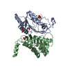
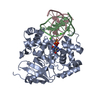
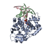
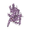
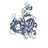
| Entry | Database: PDB / ID: 2pnq | ||||||
|---|---|---|---|---|---|---|---|
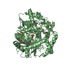
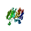
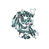
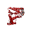
| Title | Crystal structure of pyruvate dehydrogenase phosphatase 1 (PDP1) | ||||||
 Components Components | [Pyruvate dehydrogenase [lipoamide]]-phosphatase 1 | ||||||
 Keywords Keywords |  HYDROLASE / pyruvate dehydrogenase phosphatase 1 / catalytic subunit / PDP1c HYDROLASE / pyruvate dehydrogenase phosphatase 1 / catalytic subunit / PDP1c | ||||||
| Function / homology |  Function and homology information Function and homology information[pyruvate dehydrogenase (acetyl-transferring)]-phosphatase / positive regulation of pyruvate dehydrogenase activity / Regulation of pyruvate dehydrogenase (PDH) complex / protein serine/threonine phosphatase activity => GO:0004722 / [pyruvate dehydrogenase (acetyl-transferring)]-phosphatase activity / peptidyl-threonine dephosphorylation / protein serine/threonine phosphatase activity / positive regulation of catalytic activity / protein dephosphorylation / mitochondrial matrix ...[pyruvate dehydrogenase (acetyl-transferring)]-phosphatase / positive regulation of pyruvate dehydrogenase activity / Regulation of pyruvate dehydrogenase (PDH) complex / protein serine/threonine phosphatase activity => GO:0004722 / [pyruvate dehydrogenase (acetyl-transferring)]-phosphatase activity / peptidyl-threonine dephosphorylation / protein serine/threonine phosphatase activity / positive regulation of catalytic activity / protein dephosphorylation / mitochondrial matrix / calcium ion binding / protein-containing complex binding / magnesium ion binding / mitochondrionSimilarity search - Function | ||||||
| Biological species |  Rattus norvegicus (Norway rat) Rattus norvegicus (Norway rat) | ||||||
| Method | X-RAY DIFFRACTION / SYNCHROTRON / MIR / Resolution: 1.81 Å | ||||||
 Authors Authors | Vassylyev, D.G. / Symersky, J. | ||||||
 Citation Citation | Journal: J.Mol.Biol. / Year: 2007 Title: Crystal structure of pyruvate dehydrogenase phosphatase 1 and its functional implications. Authors: Vassylyev, D.G. / Symersky, J. | ||||||
| History |
|
- Structure visualization
| Structure viewer | Molecule: MolmilJmol/JSmol |
|---|
- Downloads & links
-Download
| PDBx/mmCIF format | 2pnq.cif.gz | 175.8 KB | Display | PDBx/mmCIF format |
|---|---|---|---|---|
| PDB format | pdb2pnq.ent.gz | 137.6 KB | Display | PDB format |
| PDBx/mmJSON format | 2pnq.json.gz | Tree view | PDBx/mmJSON format | |
| Others |  Other downloads Other downloads |
-Validation report
| Arichive directory | https://data.pdbj.org/pub/pdb/validation_reports/pn/2pnqftp://data.pdbj.org/pub/pdb/validation_reports/pn/2pnq | HTTPS FTP |
|---|
-Related structure data
| Similar structure data |
|---|
-Links
 PDBj
PDBj- Assembly
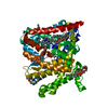
| Deposited unit | 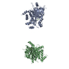
| ||||||||
|---|---|---|---|---|---|---|---|---|---|
| 1 | 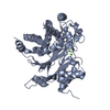
| ||||||||
| 2 | 
| ||||||||
| Unit cell |
| ||||||||
| Details | The bilogical assembly is a monomer |
-Components
| #1: Protein | Mass: 52682.223 Da / Num. of mol.: 2 Source method: isolated from a genetically manipulated source Source: (gene. exp.) Rattus norvegicus (Norway rat) / Gene: Pdp1, Ppm2c / Plasmid: pPDP1c / Species (production host): Escherichia coli / Production host:  Escherichia coli BL21(DE3) (bacteria) / Strain (production host): BL21 (DE3) Escherichia coli BL21(DE3) (bacteria) / Strain (production host): BL21 (DE3)References: UniProt: O88483, [pyruvate dehydrogenase (acetyl-transferring)]-phosphatase #2: Chemical | ChemComp-MG /   Mass: 24.305 Da / Num. of mol.: 4 / Source method: obtained synthetically / Formula: Mg Mass: 24.305 Da / Num. of mol.: 4 / Source method: obtained synthetically / Formula: Mg#3: Water | ChemComp-HOH / | Water Mass: 18.015 Da / Num. of mol.: 479 / Source method: isolated from a natural source / Formula: H2O Mass: 18.015 Da / Num. of mol.: 479 / Source method: isolated from a natural source / Formula: H2O |
|---|
-Experimental details
-Experiment
| Experiment | Method: X-RAY DIFFRACTION / Number of used crystals: 1 |
|---|
- Sample preparation
| Crystal | Density Matthews: 2.150221 Å3/Da / Density % sol: 42.79657 % |
|---|---|
| Crystal grow | Temperature: 295 K / Method: vapor diffusion, sitting drop / pH: 6.7 Details: Protein solution: protein 10mg/ml, 50 mM Tris pH 8.0, 5 mM MgCl2, 50 mM KCl, 0.1 mM EDTA Precipitant solution: 30% ethylene glycol, 0.1 M MES pH 6.0, pH 6.7, VAPOR DIFFUSION, SITTING DROP, temperature 295K |
-Data collection
| Diffraction | Mean temperature: 100 K |
|---|---|
| Diffraction source | Source: SYNCHROTRON / Site: APS  / Beamline: 22-ID / Wavelength: 1 Å / Beamline: 22-ID / Wavelength: 1 Å |
| Detector | Type: MARMOSAIC 300 mm CCD / Detector: CCD / Date: Mar 10, 2006 |
| Radiation | Monochromator: Graphite / Protocol: SINGLE WAVELENGTH / Monochromatic (M) / Laue (L): M / Scattering type: x-ray |
| Radiation wavelength | Wavelength: 1 Å / Relative weight: 1 |
| Reflection | Resolution: 1.81→30 Å / Num. all: 64759 / Num. obs: 64759 / % possible obs: 80 % / Observed criterion σ(F): 0 / Observed criterion σ(I): 0 / Redundancy: 3.3 % / Biso Wilson estimate: 40.3 Å2 / Rmerge(I) obs: 0.065 / Rsym value: 0.065 / Net I/σ(I): 15 |
| Reflection shell | Resolution: 1.81→1.87 Å / Redundancy: 2.6 % / Rmerge(I) obs: 0.48 / Mean I/σ(I) obs: 1.9 / Num. unique all: 64759 / Rsym value: 0.48 / % possible all: 52.3 |
- Processing
| Software |
| |||||||||||||||||||||||||
|---|---|---|---|---|---|---|---|---|---|---|---|---|---|---|---|---|---|---|---|---|---|---|---|---|---|---|
| Refinement | Method to determine structure: MIR Starting model: Original modeling Resolution: 1.81→30 Å / Isotropic thermal model: Isotropic / Cross valid method: THROUGHOUT / σ(F): 0 / σ(I): 0 / Stereochemistry target values: Engh & Huber Details: The diffraction images were affected by the severe ice rings whose removal resulted in a loss of ~12% of the measured reflections from the refinement. the perfect merohedral twinning was ...Details: The diffraction images were affected by the severe ice rings whose removal resulted in a loss of ~12% of the measured reflections from the refinement. the perfect merohedral twinning was detected in the crystals with the twinning operator {h,-k,-l}. the refinement statistics presented for this entry corresponds to the refinement carried out using the twinning option of the cns program. for the deposition the diffraction data were detwinned using the cns program. therefore, the refinement statistics calculated based on the detwinned data might be slightly different from those obtained during the "twinned" refinement included in this entry.
| |||||||||||||||||||||||||
| Displacement parameters | Biso mean: 40.83 Å2 | |||||||||||||||||||||||||
| Refine analyze |
| |||||||||||||||||||||||||
| Refinement step | Cycle: LAST / Resolution: 1.81→30 Å
| |||||||||||||||||||||||||
| Refine LS restraints |
| |||||||||||||||||||||||||
| LS refinement shell | Resolution: 1.81→1.87 Å
|
