 Movie
Movie Controller


+ Open data

- Basic information
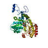
| Entry | Database: PDB / ID: 6zjf | ||||||
|---|---|---|---|---|---|---|---|
| Title | Crystal structure of STK17B (DRAK2) in complex with AP-229 | ||||||
 Components Components | Serine/threonine-protein kinase 17B | ||||||
 Keywords Keywords |  TRANSFERASE / kinase inhibitor / DRAK2 / STK17B / Structural Genomics / Structural Genomics Consortium / SGC TRANSFERASE / kinase inhibitor / DRAK2 / STK17B / Structural Genomics / Structural Genomics Consortium / SGC | ||||||
| Function / homology |  Function and homology information Function and homology informationpositive regulation of fibroblast apoptotic process / Flemming body / endoplasmic reticulum-Golgi intermediate compartment / actin cytoskeleton / protein autophosphorylation / non-specific serine/threonine protein kinase / protein kinase activity / intracellular signal transduction / positive regulation of apoptotic process / protein phosphorylation ...positive regulation of fibroblast apoptotic process / Flemming body / endoplasmic reticulum-Golgi intermediate compartment / actin cytoskeleton / protein autophosphorylation / non-specific serine/threonine protein kinase / protein kinase activity / intracellular signal transduction / positive regulation of apoptotic process / protein phosphorylation / protein serine kinase activity / protein serine/threonine kinase activity / apoptotic process / nucleoplasm / ATP binding / nucleus / plasma membraneSimilarity search - Function | ||||||
| Biological species |  Homo sapiens (human) Homo sapiens (human) | ||||||
| Method | X-RAY DIFFRACTION / SYNCHROTRON / MOLECULAR REPLACEMENT / Resolution: 1.75 Å | ||||||
 Authors Authors | Chaikuad, A. / Picado, A. / Willson, T. / Knapp, S. / Structural Genomics Consortium (SGC) | ||||||
 Citation Citation | Journal: J.Med.Chem. / Year: 2020 Title: A Chemical Probe for Dark Kinase STK17B Derives Its Potency and High Selectivity through a Unique P-Loop Conformation. Authors: Picado, A. / Chaikuad, A. / Wells, C.I. / Shrestha, S. / Zuercher, W.J. / Pickett, J.E. / Kwarcinski, F.E. / Sinha, P. / de Silva, C.S. / Zutshi, R. / Liu, S. / Kannan, N. / Knapp, S. / ...Authors: Picado, A. / Chaikuad, A. / Wells, C.I. / Shrestha, S. / Zuercher, W.J. / Pickett, J.E. / Kwarcinski, F.E. / Sinha, P. / de Silva, C.S. / Zutshi, R. / Liu, S. / Kannan, N. / Knapp, S. / Drewry, D.H. / Willson, T.M. | ||||||
| History |
|
- Structure visualization
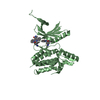
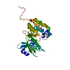
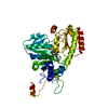
| Structure viewer | Molecule: MolmilJmol/JSmol |
|---|
- Downloads & links
-Download
| PDBx/mmCIF format | 6zjf.cif.gz | 141.2 KB | Display | PDBx/mmCIF format |
|---|---|---|---|---|
| PDB format | pdb6zjf.ent.gz | 109.5 KB | Display | PDB format |
| PDBx/mmJSON format | 6zjf.json.gz | Tree view | PDBx/mmJSON format | |
| Others |  Other downloads Other downloads |
-Validation report
| Arichive directory | https://data.pdbj.org/pub/pdb/validation_reports/zj/6zjfftp://data.pdbj.org/pub/pdb/validation_reports/zj/6zjf | HTTPS FTP |
|---|
-Related structure data
| Related structure data | 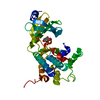 3lm5C 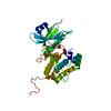 6y6fC 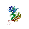 6y6hC 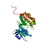 7akgC 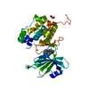 3lm0S S: Starting model for refinement C: citing same article ( |
|---|---|
| Similar structure data |


-Links
 PDBj
PDBj- Assembly
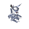
| Deposited unit | 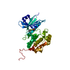
| ||||||||||||
|---|---|---|---|---|---|---|---|---|---|---|---|---|---|
| 1 |
| ||||||||||||
| Unit cell |
| ||||||||||||
| Components on special symmetry positions |
|
-Components
| #1: Protein | Mass: 37327.086 Da / Num. of mol.: 1 Source method: isolated from a genetically manipulated source Source: (gene. exp.) Homo sapiens (human) / Gene: STK17B, DRAK2 / Plasmid: pNIC28-Bsa4 / Production host:  Escherichia coli BL21(DE3) (bacteria) / Variant (production host): -R3-pRARE2 Escherichia coli BL21(DE3) (bacteria) / Variant (production host): -R3-pRARE2References: UniProt: O94768, non-specific serine/threonine protein kinase | ||||
|---|---|---|---|---|---|
| #2: Chemical | ChemComp-QM2 /   Mass: 342.435 Da / Num. of mol.: 1 / Source method: obtained synthetically / Formula: C17H14N2O2S2 / Feature type: SUBJECT OF INVESTIGATION Mass: 342.435 Da / Num. of mol.: 1 / Source method: obtained synthetically / Formula: C17H14N2O2S2 / Feature type: SUBJECT OF INVESTIGATION | ||||
| #3: Chemical | Ethylene glycol  Mass: 62.068 Da / Num. of mol.: 2 / Source method: obtained synthetically / Formula: C2H6O2 Mass: 62.068 Da / Num. of mol.: 2 / Source method: obtained synthetically / Formula: C2H6O2#4: Water | ChemComp-HOH / | Water Mass: 18.015 Da / Num. of mol.: 231 / Source method: isolated from a natural source / Formula: H2O Mass: 18.015 Da / Num. of mol.: 231 / Source method: isolated from a natural source / Formula: H2OHas ligand of interest | Y | |
-Experimental details
-Experiment
| Experiment | Method: X-RAY DIFFRACTION / Number of used crystals: 1 |
|---|
- Sample preparation
| Crystal | Density Matthews: 2.66 Å3/Da / Density % sol: 53.73 % |
|---|---|
| Crystal grow | Temperature: 277.15 K / Method: vapor diffusion, sitting drop / pH: 6 / Details: 19% PEG 3350, 0.1M NaCl, 0.1M bis-tris 6.0 |
-Data collection
| Diffraction | Mean temperature: 100 K / Serial crystal experiment: N | |||||||||||||||||||||||||||
|---|---|---|---|---|---|---|---|---|---|---|---|---|---|---|---|---|---|---|---|---|---|---|---|---|---|---|---|---|
| Diffraction source | Source: SYNCHROTRON / Site: PETRA III, EMBL c/o DESY  / Beamline: P13 (MX1) / Wavelength: 1.0332 Å / Beamline: P13 (MX1) / Wavelength: 1.0332 Å | |||||||||||||||||||||||||||
| Detector | Type: DECTRIS PILATUS3 6M / Detector: PIXEL / Date: Nov 27, 2018 | |||||||||||||||||||||||||||
| Radiation | Protocol: SINGLE WAVELENGTH / Monochromatic (M) / Laue (L): M / Scattering type: x-ray | |||||||||||||||||||||||||||
| Radiation wavelength | Wavelength: 1.0332 Å / Relative weight: 1 | |||||||||||||||||||||||||||
| Reflection | Resolution: 1.75→37.27 Å / Num. obs: 41311 / % possible obs: 100 % / Redundancy: 7.2 % / CC1/2: 0.998 / Rmerge(I) obs: 0.084 / Rpim(I) all: 0.036 / Rrim(I) all: 0.096 / Net I/σ(I): 11.9 | |||||||||||||||||||||||||||
| Reflection shell | Diffraction-ID: 1
|
- Processing
| Software |
| ||||||||||||||||||||||||||||||||||||||||||||||||||||||||||||
|---|---|---|---|---|---|---|---|---|---|---|---|---|---|---|---|---|---|---|---|---|---|---|---|---|---|---|---|---|---|---|---|---|---|---|---|---|---|---|---|---|---|---|---|---|---|---|---|---|---|---|---|---|---|---|---|---|---|---|---|---|---|
| Refinement | Method to determine structure: MOLECULAR REPLACEMENT Starting model: 3LM0 Resolution: 1.75→37.27 Å / Cor.coef. Fo:Fc: 0.962 / Cor.coef. Fo:Fc free: 0.945 / SU B: 4.68 / SU ML: 0.073 / SU R Cruickshank DPI: 0.0996 / Cross valid method: THROUGHOUT / σ(F): 0 / ESU R: 0.1 / ESU R Free: 0.099 / Stereochemistry target values: MAXIMUM LIKELIHOOD Details: U VALUES : WITH TLS ADDED HYDROGENS HAVE BEEN ADDED IN THE RIDING POSITIONS
| ||||||||||||||||||||||||||||||||||||||||||||||||||||||||||||
| Solvent computation | Ion probe radii: 0.8 Å / Shrinkage radii: 0.8 Å / VDW probe radii: 1.2 Å / Solvent model: MASK | ||||||||||||||||||||||||||||||||||||||||||||||||||||||||||||
| Displacement parameters | Biso max: 108.56 Å2 / Biso mean: 31.099 Å2 / Biso min: 15.82 Å2
| ||||||||||||||||||||||||||||||||||||||||||||||||||||||||||||
| Refinement step | Cycle: final / Resolution: 1.75→37.27 Å
| ||||||||||||||||||||||||||||||||||||||||||||||||||||||||||||
| Refine LS restraints |
| ||||||||||||||||||||||||||||||||||||||||||||||||||||||||||||
| LS refinement shell | Resolution: 1.75→1.795 Å / Rfactor Rfree error: 0 / Total num. of bins used: 20
| ||||||||||||||||||||||||||||||||||||||||||||||||||||||||||||
| Refinement TLS params. | Method: refined / Origin x: 60.1676 Å / Origin y: 11.9419 Å / Origin z: -5.3187 Å
|
