 Movie
Movie Controller

[English] 日本語
 Yorodumi
- PDB-3lm5: Crystal Structure of human Serine/Threonine Kinase 17B (STK17B) i... -
+ Open data

- Basic information
| Entry | Database: PDB / ID: 3lm5 | ||||||
|---|---|---|---|---|---|---|---|
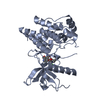
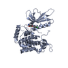
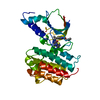
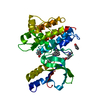
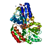
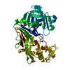
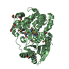
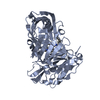
| Title | Crystal Structure of human Serine/Threonine Kinase 17B (STK17B) in complex with Quercetin | ||||||
 Components Components | Serine/threonine-protein kinase 17B | ||||||
 Keywords Keywords |  TRANSFERASE / STK17B / serine/threonine kinase 17B / DRAK2 / DAP kinase related apoptosis-inducing protein kinase 2 / death-associated protein kinase-related 2 / Structural Genomics / Structural Genomics Consortium / SGC / Apoptosis / ATP-binding / Kinase / Nucleotide-binding / Nucleus / Phosphoprotein / Serine/threonine-protein kinase TRANSFERASE / STK17B / serine/threonine kinase 17B / DRAK2 / DAP kinase related apoptosis-inducing protein kinase 2 / death-associated protein kinase-related 2 / Structural Genomics / Structural Genomics Consortium / SGC / Apoptosis / ATP-binding / Kinase / Nucleotide-binding / Nucleus / Phosphoprotein / Serine/threonine-protein kinase | ||||||
| Function / homology |  Function and homology information Function and homology informationpositive regulation of fibroblast apoptotic process / Flemming body / endoplasmic reticulum-Golgi intermediate compartment / actin cytoskeleton / protein autophosphorylation / non-specific serine/threonine protein kinase / protein kinase activity / intracellular signal transduction / positive regulation of apoptotic process / protein phosphorylation ...positive regulation of fibroblast apoptotic process / Flemming body / endoplasmic reticulum-Golgi intermediate compartment / actin cytoskeleton / protein autophosphorylation / non-specific serine/threonine protein kinase / protein kinase activity / intracellular signal transduction / positive regulation of apoptotic process / protein phosphorylation / protein serine kinase activity / protein serine/threonine kinase activity / apoptotic process / nucleoplasm / ATP binding / nucleus / plasma membraneSimilarity search - Function | ||||||
| Biological species |  Homo sapiens (human) Homo sapiens (human) | ||||||
| Method | X-RAY DIFFRACTION / SYNCHROTRON / MOLECULAR REPLACEMENT / Resolution: 2.29 Å | ||||||
 Authors Authors | Ugochukwu, E. / Soundararajan, M. / Rellos, P. / Fedorov, O. / Phillips, C. / Wang, J. / Hapka, E. / Filippakopoulos, P. / Chaikuad, A. / Pike, A.C.W. ...Ugochukwu, E. / Soundararajan, M. / Rellos, P. / Fedorov, O. / Phillips, C. / Wang, J. / Hapka, E. / Filippakopoulos, P. / Chaikuad, A. / Pike, A.C.W. / Carpenter, L. / Vollmar, M. / von Delft, F. / Bountra, C. / Arrowsmith, C.H. / Weigelt, J. / Edwards, A. / Knapp, S. / Structural Genomics Consortium (SGC) | ||||||
 Citation Citation | Journal: J.Med.Chem. / Year: 2020 Title: A Chemical Probe for Dark Kinase STK17B Derives Its Potency and High Selectivity through a Unique P-Loop Conformation. Authors: Picado, A. / Chaikuad, A. / Wells, C.I. / Shrestha, S. / Zuercher, W.J. / Pickett, J.E. / Kwarcinski, F.E. / Sinha, P. / de Silva, C.S. / Zutshi, R. / Liu, S. / Kannan, N. / Knapp, S. / ...Authors: Picado, A. / Chaikuad, A. / Wells, C.I. / Shrestha, S. / Zuercher, W.J. / Pickett, J.E. / Kwarcinski, F.E. / Sinha, P. / de Silva, C.S. / Zutshi, R. / Liu, S. / Kannan, N. / Knapp, S. / Drewry, D.H. / Willson, T.M. | ||||||
| History |
|
- Structure visualization
| Structure viewer | Molecule: MolmilJmol/JSmol |
|---|
- Downloads & links
-Download
| PDBx/mmCIF format | 3lm5.cif.gz | 74 KB | Display | PDBx/mmCIF format |
|---|---|---|---|---|
| PDB format | pdb3lm5.ent.gz | 53.4 KB | Display | PDB format |
| PDBx/mmJSON format | 3lm5.json.gz | Tree view | PDBx/mmJSON format | |
| Others |  Other downloads Other downloads |
-Validation report
| Arichive directory | https://data.pdbj.org/pub/pdb/validation_reports/lm/3lm5ftp://data.pdbj.org/pub/pdb/validation_reports/lm/3lm5 | HTTPS FTP |
|---|
-Related structure data
| Related structure data | 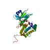 6y6fC 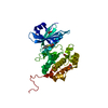 6y6hC 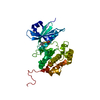 6zjfC 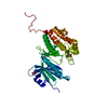 7akgC  3ehaS S: Starting model for refinement C: citing same article ( |
|---|---|
| Similar structure data |


-Links
 PDBj
PDBj
- Assembly
| Deposited unit | 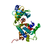
| ||||||||
|---|---|---|---|---|---|---|---|---|---|
| 1 |
| ||||||||
| Unit cell |
|
-Components
| #1: Protein | Mass: 37327.086 Da / Num. of mol.: 1 / Fragment: STK17B Source method: isolated from a genetically manipulated source Source: (gene. exp.) Homo sapiens (human) / Gene: DRAK2, STK17B / Plasmid: pNIC28-Bsa4 / Production host:  Escherichia coli (E. coli) / Strain (production host): BL21(DE3)-R3-pRARE2 Escherichia coli (E. coli) / Strain (production host): BL21(DE3)-R3-pRARE2References: UniProt: O94768, non-specific serine/threonine protein kinase |
|---|---|
| #2: Chemical | ChemComp-QUE / Quercetin  Mass: 302.236 Da / Num. of mol.: 1 / Source method: obtained synthetically / Formula: C15H10O7 Mass: 302.236 Da / Num. of mol.: 1 / Source method: obtained synthetically / Formula: C15H10O7 |
| #3: Water | ChemComp-HOH / Water Mass: 18.015 Da / Num. of mol.: 118 / Source method: isolated from a natural source / Formula: H2O Mass: 18.015 Da / Num. of mol.: 118 / Source method: isolated from a natural source / Formula: H2O |
-Experimental details
-Experiment
| Experiment | Method: X-RAY DIFFRACTION / Number of used crystals: 1 |
|---|
- Sample preparation
| Crystal | Density Matthews: 2.68 Å3/Da / Density % sol: 54.18 % |
|---|---|
| Crystal grow | Temperature: 277 K / Method: vapor diffusion, sitting drop / pH: 5.5 Details: 0.2M NaCl; 0.1M BIS-TRIS pH 5.5; 25% PEG 3350, VAPOR DIFFUSION, SITTING DROP, temperature 277K |
-Data collection
| Diffraction | Mean temperature: 100 K |
|---|---|
| Diffraction source | Source: SYNCHROTRON / Site: Diamond  / Beamline: I02 / Wavelength: 0.98 Å / Beamline: I02 / Wavelength: 0.98 Å |
| Detector | Type: MAR225 / Detector: CCD / Date: Dec 17, 2009 |
| Radiation | Protocol: SINGLE WAVELENGTH / Monochromatic (M) / Laue (L): M / Scattering type: x-ray |
| Radiation wavelength | Wavelength: 0.98 Å / Relative weight: 1 |
| Reflection | Resolution: 2.29→52.49 Å / Num. obs: 18152 / % possible obs: 93.2 % / Observed criterion σ(F): 0 / Observed criterion σ(I): 0 / Redundancy: 6.6 % / Rmerge(I) obs: 0.097 / Rsym value: 0.097 / Net I/σ(I): 12.5 |
| Reflection shell | Resolution: 2.29→2.39 Å / Redundancy: 6.9 % / Rmerge(I) obs: 0.902 / Mean I/σ(I) obs: 2.2 / Rsym value: 0.902 / % possible all: 100 |
- Processing
| Software |
| ||||||||||||||||||||||||||||||||||||||||||||||||||||||||||||||||||||||||||||||||||||||||||||||||||||||||||||||||||||||||||||||||||||||||||||||||||||||||||||||||||||||||||
|---|---|---|---|---|---|---|---|---|---|---|---|---|---|---|---|---|---|---|---|---|---|---|---|---|---|---|---|---|---|---|---|---|---|---|---|---|---|---|---|---|---|---|---|---|---|---|---|---|---|---|---|---|---|---|---|---|---|---|---|---|---|---|---|---|---|---|---|---|---|---|---|---|---|---|---|---|---|---|---|---|---|---|---|---|---|---|---|---|---|---|---|---|---|---|---|---|---|---|---|---|---|---|---|---|---|---|---|---|---|---|---|---|---|---|---|---|---|---|---|---|---|---|---|---|---|---|---|---|---|---|---|---|---|---|---|---|---|---|---|---|---|---|---|---|---|---|---|---|---|---|---|---|---|---|---|---|---|---|---|---|---|---|---|---|---|---|---|---|---|---|---|
| Refinement | Method to determine structure: MOLECULAR REPLACEMENT Starting model: PDB entry 3EHA Resolution: 2.29→52.49 Å / Cor.coef. Fo:Fc: 0.946 / Cor.coef. Fo:Fc free: 0.906 / SU B: 13.051 / SU ML: 0.15 / TLS residual ADP flag: LIKELY RESIDUAL / Cross valid method: THROUGHOUT / ESU R: 0.271 / ESU R Free: 0.252 / Stereochemistry target values: MAXIMUM LIKELIHOOD / Details: HYDROGENS HAVE BEEN ADDED IN THE RIDING POSITIONS
| ||||||||||||||||||||||||||||||||||||||||||||||||||||||||||||||||||||||||||||||||||||||||||||||||||||||||||||||||||||||||||||||||||||||||||||||||||||||||||||||||||||||||||
| Solvent computation | Ion probe radii: 0.8 Å / Shrinkage radii: 0.8 Å / VDW probe radii: 1.4 Å / Solvent model: MASK | ||||||||||||||||||||||||||||||||||||||||||||||||||||||||||||||||||||||||||||||||||||||||||||||||||||||||||||||||||||||||||||||||||||||||||||||||||||||||||||||||||||||||||
| Displacement parameters | Biso mean: 18.239 Å2
| ||||||||||||||||||||||||||||||||||||||||||||||||||||||||||||||||||||||||||||||||||||||||||||||||||||||||||||||||||||||||||||||||||||||||||||||||||||||||||||||||||||||||||
| Refinement step | Cycle: LAST / Resolution: 2.29→52.49 Å
| ||||||||||||||||||||||||||||||||||||||||||||||||||||||||||||||||||||||||||||||||||||||||||||||||||||||||||||||||||||||||||||||||||||||||||||||||||||||||||||||||||||||||||
| Refine LS restraints |
| ||||||||||||||||||||||||||||||||||||||||||||||||||||||||||||||||||||||||||||||||||||||||||||||||||||||||||||||||||||||||||||||||||||||||||||||||||||||||||||||||||||||||||
| LS refinement shell | Resolution: 2.285→2.345 Å / Total num. of bins used: 20
| ||||||||||||||||||||||||||||||||||||||||||||||||||||||||||||||||||||||||||||||||||||||||||||||||||||||||||||||||||||||||||||||||||||||||||||||||||||||||||||||||||||||||||
| Refinement TLS params. | Method: refined / Refine-ID: X-RAY DIFFRACTION
| ||||||||||||||||||||||||||||||||||||||||||||||||||||||||||||||||||||||||||||||||||||||||||||||||||||||||||||||||||||||||||||||||||||||||||||||||||||||||||||||||||||||||||
| Refinement TLS group |
|
