 Movie
Movie Controller

[English] 日本語
 Yorodumi
- PDB-6ww0: Human steroidogenic cytochrome P450 17A1 with 3-keto-5alpha-abira... -
+ Open data

- Basic information
| Entry | Database: PDB / ID: 6ww0 | ||||||
|---|---|---|---|---|---|---|---|
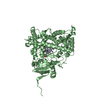
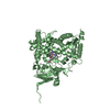
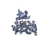
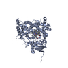
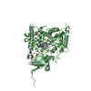
| Title | Human steroidogenic cytochrome P450 17A1 with 3-keto-5alpha-abiraterone analog | ||||||
 Components Components | Steroid 17-alpha-hydroxylase/17,20 lyase | ||||||
 Keywords Keywords |  OXIDOREDUCTASE / Cytochrome P450 / P450 / 17A1 OXIDOREDUCTASE / Cytochrome P450 / P450 / 17A1 | ||||||
| Function / homology |  Function and homology information Function and homology informationDefective CYP17A1 causes AH5 / steroid 17alpha-monooxygenase / 17alpha-hydroxyprogesterone deacetylase / steroid 17-alpha-monooxygenase activity / : / glucocorticoid biosynthetic process / Androgen biosynthesis / hormone biosynthetic process / Glucocorticoid biosynthesis / androgen biosynthetic process ...Defective CYP17A1 causes AH5 / steroid 17alpha-monooxygenase / 17alpha-hydroxyprogesterone deacetylase / steroid 17-alpha-monooxygenase activity / : / glucocorticoid biosynthetic process / Androgen biosynthesis / hormone biosynthetic process / Glucocorticoid biosynthesis / androgen biosynthetic process / sex differentiation / progesterone metabolic process / steroid biosynthetic process / steroid metabolic process / oxygen binding / iron ion binding / axon / neuronal cell body / heme binding / endoplasmic reticulum membrane / endoplasmic reticulumSimilarity search - Function | ||||||
| Biological species |  Homo sapiens (human) Homo sapiens (human) | ||||||
| Method | X-RAY DIFFRACTION / SYNCHROTRON / MOLECULAR REPLACEMENT / Resolution: 2.01 Å | ||||||
 Authors Authors | Petrunak, E.M. / Bart, A.G. / Scott, E.E. | ||||||
| Funding support |  United States, 1items United States, 1items
| ||||||
 Citation Citation | Journal: J.Biol.Chem. / Year: 2023 Title: Human cytochrome P450 17A1 structures with metabolites of prostate cancer drug abiraterone reveal substrate-binding plasticity and a second binding site. Authors: Petrunak, E.M. / Bart, A.G. / Peng, H.M. / Auchus, R.J. / Scott, E.E. | ||||||
| History |
|
- Structure visualization
| Structure viewer | Molecule: MolmilJmol/JSmol |
|---|
- Downloads & links
-Download
| PDBx/mmCIF format | 6ww0.cif.gz | 786.1 KB | Display | PDBx/mmCIF format |
|---|---|---|---|---|
| PDB format | pdb6ww0.ent.gz | 609.7 KB | Display | PDB format |
| PDBx/mmJSON format | 6ww0.json.gz | Tree view | PDBx/mmJSON format | |
| Others |  Other downloads Other downloads |
-Validation report
| Arichive directory | https://data.pdbj.org/pub/pdb/validation_reports/ww/6ww0ftp://data.pdbj.org/pub/pdb/validation_reports/ww/6ww0 | HTTPS FTP |
|---|
-Related structure data
| Related structure data | 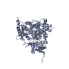 5uysC  6wr0C 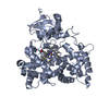 6wr1C 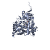 5irqS S: Starting model for refinement C: citing same article ( |
|---|---|
| Similar structure data |


-Links
 PDBj
PDBj

- Assembly
| Deposited unit | 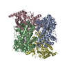
| ||||||||||||||||||||||||||||||||||||||||||||||||||||||||||||||
|---|---|---|---|---|---|---|---|---|---|---|---|---|---|---|---|---|---|---|---|---|---|---|---|---|---|---|---|---|---|---|---|---|---|---|---|---|---|---|---|---|---|---|---|---|---|---|---|---|---|---|---|---|---|---|---|---|---|---|---|---|---|---|---|
| 1 | 
| ||||||||||||||||||||||||||||||||||||||||||||||||||||||||||||||
| 2 | 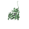
| ||||||||||||||||||||||||||||||||||||||||||||||||||||||||||||||
| 3 | 
| ||||||||||||||||||||||||||||||||||||||||||||||||||||||||||||||
| 4 | 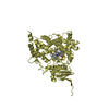
| ||||||||||||||||||||||||||||||||||||||||||||||||||||||||||||||
| Unit cell |
| ||||||||||||||||||||||||||||||||||||||||||||||||||||||||||||||
| Noncrystallographic symmetry (NCS) | NCS domain:
NCS domain segments:
|