 Movie
Movie Controller

[English] 日本語
 Yorodumi
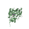
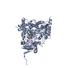
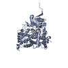
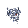
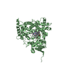
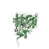
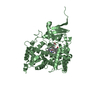
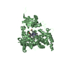
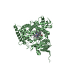
- PDB-6cir: Human Cytochrome P450 17A1 in complex with inhibitor: abiraterone... -
+ Open data

- Basic information
| Entry | Database: PDB / ID: 6cir | ||||||
|---|---|---|---|---|---|---|---|
| Title | Human Cytochrome P450 17A1 in complex with inhibitor: abiraterone C6 oxime | ||||||
 Components Components | Steroid 17-alpha-hydroxylase/17,20 lyase | ||||||
 Keywords Keywords | OXIDOREDUCTASE/OXIDOREDUCTASE INHIBITOR /  CYTOCHROME P450 / P450 / CYP17A1 / P450C17 / P450 17A1 / MONOOXYGENASE / 17A-HYDROXYLASE / 17 / 20-LYASE / HEME PROTEIN / CYTOCHROME P450 OXIDOREDUCTASE / MEMBRANE / MICROSOME / ENDOPLASMIC RETICULUM / OXIDOREDUCTASE-OXIDOREDUCTASE INHIBITOR / OXIDOREDUCTASE / OXIDOREDUCTASE-OXIDOREDUCTASE INHIBITOR complex CYTOCHROME P450 / P450 / CYP17A1 / P450C17 / P450 17A1 / MONOOXYGENASE / 17A-HYDROXYLASE / 17 / 20-LYASE / HEME PROTEIN / CYTOCHROME P450 OXIDOREDUCTASE / MEMBRANE / MICROSOME / ENDOPLASMIC RETICULUM / OXIDOREDUCTASE-OXIDOREDUCTASE INHIBITOR / OXIDOREDUCTASE / OXIDOREDUCTASE-OXIDOREDUCTASE INHIBITOR complex | ||||||
| Function / homology |  Function and homology information Function and homology informationDefective CYP17A1 causes AH5 / steroid 17alpha-monooxygenase / 17alpha-hydroxyprogesterone deacetylase / steroid 17-alpha-monooxygenase activity / : / glucocorticoid biosynthetic process / Androgen biosynthesis / hormone biosynthetic process / Glucocorticoid biosynthesis / androgen biosynthetic process ...Defective CYP17A1 causes AH5 / steroid 17alpha-monooxygenase / 17alpha-hydroxyprogesterone deacetylase / steroid 17-alpha-monooxygenase activity / : / glucocorticoid biosynthetic process / Androgen biosynthesis / hormone biosynthetic process / Glucocorticoid biosynthesis / androgen biosynthetic process / sex differentiation / progesterone metabolic process / steroid biosynthetic process / steroid metabolic process / oxygen binding / iron ion binding / axon / neuronal cell body / heme binding / endoplasmic reticulum membrane / endoplasmic reticulumSimilarity search - Function | ||||||
| Biological species |  Homo sapiens (human) Homo sapiens (human) | ||||||
| Method | X-RAY DIFFRACTION / SYNCHROTRON / MOLECULAR REPLACEMENT / Resolution: 2.648 Å | ||||||
 Authors Authors | Scott, E.E. / Fehl, C. | ||||||
| Funding support |  United States, 1items United States, 1items
| ||||||
 Citation Citation | Journal: J. Med. Chem. / Year: 2018 Title: Structure-Based Design of Inhibitors with Improved Selectivity for Steroidogenic Cytochrome P450 17A1 over Cytochrome P450 21A2. Authors: Fehl, C. / Vogt, C.D. / Yadav, R. / Li, K. / Scott, E.E. / Aube, J. | ||||||
| History |
|
- Structure visualization
| Structure viewer | Molecule: MolmilJmol/JSmol |
|---|
- Downloads & links
-Download
| PDBx/mmCIF format | 6cir.cif.gz | 704.6 KB | Display | PDBx/mmCIF format |
|---|---|---|---|---|
| PDB format | pdb6cir.ent.gz | 593.2 KB | Display | PDB format |
| PDBx/mmJSON format | 6cir.json.gz | Tree view | PDBx/mmJSON format | |
| Others |  Other downloads Other downloads |
-Validation report
| Arichive directory | https://data.pdbj.org/pub/pdb/validation_reports/ci/6cirftp://data.pdbj.org/pub/pdb/validation_reports/ci/6cir | HTTPS FTP |
|---|
-Related structure data
| Related structure data |  6chiC  6cizC  3swzS S: Starting model for refinement C: citing same article ( |
|---|---|
| Similar structure data |
-Links
 PDBj
PDBj

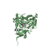
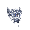
- Assembly
| Deposited unit | 
| ||||||||
|---|---|---|---|---|---|---|---|---|---|
| 1 | 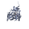
| ||||||||
| 2 | 
| ||||||||
| 3 | 
| ||||||||
| 4 | 
| ||||||||
| Unit cell |
|
-Components
| #1: Protein | Mass: 55740.141 Da / Num. of mol.: 4 / Fragment: residues 24-508 Source method: isolated from a genetically manipulated source Source: (gene. exp.) Homo sapiens (human) / Gene: CYP17A1, CYP17, S17AH / Production host:  Escherichia coli (E. coli) Escherichia coli (E. coli)References: UniProt: P05093, steroid 17alpha-monooxygenase, 17alpha-hydroxyprogesterone deacetylase#2: Chemical | ChemComp-HEM / Heme B  Mass: 616.487 Da / Num. of mol.: 4 / Source method: obtained synthetically / Formula: C34H32FeN4O4 Mass: 616.487 Da / Num. of mol.: 4 / Source method: obtained synthetically / Formula: C34H32FeN4O4#3: Chemical | ChemComp-3NQ /   Mass: 380.523 Da / Num. of mol.: 4 / Source method: obtained synthetically / Formula: C24H32N2O2 Mass: 380.523 Da / Num. of mol.: 4 / Source method: obtained synthetically / Formula: C24H32N2O2#4: Water | ChemComp-HOH / | Water Mass: 18.015 Da / Num. of mol.: 133 / Source method: isolated from a natural source / Formula: H2O Mass: 18.015 Da / Num. of mol.: 133 / Source method: isolated from a natural source / Formula: H2O |
|---|
-Experimental details
-Experiment
| Experiment | Method: X-RAY DIFFRACTION / Number of used crystals: 1 |
|---|
- Sample preparation
| Crystal | Density Matthews: 2.63 Å3/Da / Density % sol: 53.27 % |
|---|---|
| Crystal grow | Temperature: 293 K / Method: vapor diffusion, hanging drop / pH: 8.5 Details: 0.175 M Tris, pH 8.5 containing 30% PEG 3350, 3% glycerol, and 0.250-0.275 M lithium sulfate |
-Data collection
| Diffraction | Mean temperature: 100 K |
|---|---|
| Diffraction source | Source: SYNCHROTRON / Site: SSRL / Beamline: BL12-2 / Wavelength: 0.9795 Å |
| Detector | Type: DECTRIS PILATUS 6M / Detector: PIXEL / Date: Feb 17, 2014 |
| Radiation | Protocol: SINGLE WAVELENGTH / Monochromatic (M) / Laue (L): M / Scattering type: x-ray |
| Radiation wavelength | Wavelength: 0.9795 Å / Relative weight: 1 |
| Reflection | Resolution: 2.648→39.314 Å / Num. obs: 68518 / % possible obs: 99 % / Redundancy: 6.7 % / Rpim(I) all: 0.061 / Net I/σ(I): 13.1 |
| Reflection shell | Resolution: 2.65→2.79 Å / Redundancy: 6.7 % / Mean I/σ(I) obs: 1.6 / Num. unique obs: 9510 / Rpim(I) all: 0.552 / % possible all: 95.4 |
- Processing
| Software |
| ||||||||||||||||||||||||||||||||||||||||||||||||||||||||||||||||||||||||||||||||||||||||||||||||||||||||||||||||||||||||||||||||||||||||||||||||||||||||||||||||||||||||||||||||||||||
|---|---|---|---|---|---|---|---|---|---|---|---|---|---|---|---|---|---|---|---|---|---|---|---|---|---|---|---|---|---|---|---|---|---|---|---|---|---|---|---|---|---|---|---|---|---|---|---|---|---|---|---|---|---|---|---|---|---|---|---|---|---|---|---|---|---|---|---|---|---|---|---|---|---|---|---|---|---|---|---|---|---|---|---|---|---|---|---|---|---|---|---|---|---|---|---|---|---|---|---|---|---|---|---|---|---|---|---|---|---|---|---|---|---|---|---|---|---|---|---|---|---|---|---|---|---|---|---|---|---|---|---|---|---|---|---|---|---|---|---|---|---|---|---|---|---|---|---|---|---|---|---|---|---|---|---|---|---|---|---|---|---|---|---|---|---|---|---|---|---|---|---|---|---|---|---|---|---|---|---|---|---|---|---|
| Refinement | Method to determine structure: MOLECULAR REPLACEMENT Starting model: 3SWZ Resolution: 2.648→39.314 Å / SU ML: 0.39 / Cross valid method: FREE R-VALUE / σ(F): 1.34 / Phase error: 25.6 / Stereochemistry target values: ML
| ||||||||||||||||||||||||||||||||||||||||||||||||||||||||||||||||||||||||||||||||||||||||||||||||||||||||||||||||||||||||||||||||||||||||||||||||||||||||||||||||||||||||||||||||||||||
| Solvent computation | Shrinkage radii: 0.9 Å / VDW probe radii: 1.11 Å / Solvent model: FLAT BULK SOLVENT MODEL | ||||||||||||||||||||||||||||||||||||||||||||||||||||||||||||||||||||||||||||||||||||||||||||||||||||||||||||||||||||||||||||||||||||||||||||||||||||||||||||||||||||||||||||||||||||||
| Refinement step | Cycle: LAST / Resolution: 2.648→39.314 Å
| ||||||||||||||||||||||||||||||||||||||||||||||||||||||||||||||||||||||||||||||||||||||||||||||||||||||||||||||||||||||||||||||||||||||||||||||||||||||||||||||||||||||||||||||||||||||
| Refine LS restraints |
| ||||||||||||||||||||||||||||||||||||||||||||||||||||||||||||||||||||||||||||||||||||||||||||||||||||||||||||||||||||||||||||||||||||||||||||||||||||||||||||||||||||||||||||||||||||||
| LS refinement shell |
|
