 Movie
Movie Controller


+ Open data

- Basic information
| Entry | Database: PDB / ID: 6ns1 | |||||||||
|---|---|---|---|---|---|---|---|---|---|---|
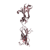
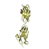
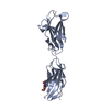
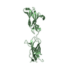
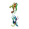
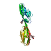
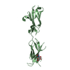
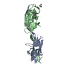
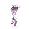
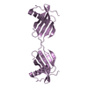
| Title | Crystal structure of DIP-gamma IG1+IG2 | |||||||||
 Components Components | Dpr-interacting protein gamma | |||||||||
 Keywords Keywords |  CELL ADHESION / Immunoglobulin superfamily / Glycoprotein / Neuronal / Cell surface receptor CELL ADHESION / Immunoglobulin superfamily / Glycoprotein / Neuronal / Cell surface receptor | |||||||||
| Function / homology |  Function and homology information Function and homology informationregulation of neuromuscular junction development / Role of ABL in ROBO-SLIT signaling / Signaling by ROBO receptors / : / Regulation of expression of SLITs and ROBOs / neuron projection membrane / photoreceptor cell axon guidance / establishment of synaptic specificity at neuromuscular junction / synapse organization / neuron projection / plasma membraneSimilarity search - Function | |||||||||
| Biological species |  Drosophila melanogaster (fruit fly) Drosophila melanogaster (fruit fly) | |||||||||
| Method | X-RAY DIFFRACTION / SYNCHROTRON / MOLECULAR REPLACEMENT / Resolution: 1.85 Å | |||||||||
 Authors Authors | Cheng, S. / Park, Y.J. / Kurleto, J.D. / Ozkan, E. | |||||||||
| Funding support |  United States, 1items United States, 1items
| |||||||||
 Citation Citation | Journal: Elife / Year: 2019 Title: Molecular basis of synaptic specificity by immunoglobulin superfamily receptors in Drosophila. Authors: Cheng, S. / Ashley, J. / Kurleto, J.D. / Lobb-Rabe, M. / Park, Y.J. / Carrillo, R.A. / Ozkan, E. | |||||||||
| History |
|
- Structure visualization
| Structure viewer | Molecule: MolmilJmol/JSmol |
|---|
- Downloads & links
-Download
| PDBx/mmCIF format | 6ns1.cif.gz | 63.2 KB | Display | PDBx/mmCIF format |
|---|---|---|---|---|
| PDB format | pdb6ns1.ent.gz | 43 KB | Display | PDB format |
| PDBx/mmJSON format | 6ns1.json.gz | Tree view | PDBx/mmJSON format | |
| Others |  Other downloads Other downloads |
-Validation report
| Arichive directory | https://data.pdbj.org/pub/pdb/validation_reports/ns/6ns1ftp://data.pdbj.org/pub/pdb/validation_reports/ns/6ns1 | HTTPS FTP |
|---|
-Related structure data
| Related structure data | 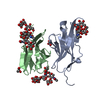 6nrqC 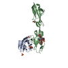 6nrrC 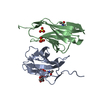 6nrwC  6nrxC 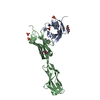 5eo9S S: Starting model for refinement C: citing same article ( |
|---|---|
| Similar structure data |
-Links
 PDBj
PDBj


- Assembly
| Deposited unit | 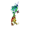
| ||||||||
|---|---|---|---|---|---|---|---|---|---|
| 1 |
| ||||||||
| Unit cell |
|
-Components
| #1: Protein | Mass: 24006.553 Da / Num. of mol.: 1 Source method: isolated from a genetically manipulated source Source: (gene. exp.) Drosophila melanogaster (fruit fly)Gene: DIP-gamma, 14521, anon-WO0140519.196, CT34248, Dmel\CG14521, CG14521, Dmel_CG14521 Cell line (production host): High Five cells / Production host: Trichoplusia ni (cabbage looper) / References: UniProt: Q9VAR6 |
|---|---|
| #2: Polysaccharide | 2-acetamido-2-deoxy-beta-D-glucopyranose-(1-4)-[alpha-L-fucopyranose-(1-6)]2-acetamido-2-deoxy-beta- ...2-acetamido-2-deoxy-beta-D-glucopyranose-(1-4)-[alpha-L-fucopyranose-(1-6)]2-acetamido-2-deoxy-beta-D-glucopyranose / Mass: 570.542 Da / Num. of mol.: 1 Source method: isolated from a genetically manipulated source |
| #3: Water | ChemComp-HOH / Water Mass: 18.015 Da / Num. of mol.: 179 / Source method: isolated from a natural source / Formula: H2O Mass: 18.015 Da / Num. of mol.: 179 / Source method: isolated from a natural source / Formula: H2O |
-Experimental details
-Experiment
| Experiment | Method: X-RAY DIFFRACTION / Number of used crystals: 1 |
|---|
- Sample preparation
| Crystal | Density Matthews: 2.29 Å3/Da / Density % sol: 46.19 % |
|---|---|
| Crystal grow | Temperature: 294 K / Method: vapor diffusion, sitting drop / pH: 5.5 Details: 0.15 M ammonium sulfate, 0.1 M MES, pH 5.5, 25% (w/v) PEG 4000 |
-Data collection
| Diffraction | Mean temperature: 120 K / Serial crystal experiment: N |
|---|---|
| Diffraction source | Source: SYNCHROTRON / Site: APS / Beamline: 24-ID-E / Wavelength: 0.97919 Å |
| Detector | Type: ADSC QUANTUM 315 / Detector: CCD / Date: Nov 24, 2015 |
| Radiation | Monochromator: Cryogenically-cooled single crystal Si(220) / Protocol: SINGLE WAVELENGTH / Monochromatic (M) / Laue (L): M / Scattering type: x-ray |
| Radiation wavelength | Wavelength: 0.97919 Å / Relative weight: 1 |
| Reflection | Resolution: 1.85→50 Å / Num. obs: 18742 / % possible obs: 99.9 % / Observed criterion σ(I): -3 / Redundancy: 6.32 % / Biso Wilson estimate: 26.72 Å2 / CC1/2: 0.996 / Rmerge(I) obs: 0.146 / Rrim(I) all: 0.158 / Net I/σ(I): 8.81 |
| Reflection shell | Resolution: 1.85→1.9 Å / Redundancy: 3.79 % / Rmerge(I) obs: 0.716 / Mean I/σ(I) obs: 1.6 / Num. unique obs: 1366 / CC1/2: 0.828 / Rrim(I) all: 0.833 / % possible all: 99.8 |
- Processing
| Software |
| ||||||||||||||||||||||||||||||||||||||||||||||||||||||||
|---|---|---|---|---|---|---|---|---|---|---|---|---|---|---|---|---|---|---|---|---|---|---|---|---|---|---|---|---|---|---|---|---|---|---|---|---|---|---|---|---|---|---|---|---|---|---|---|---|---|---|---|---|---|---|---|---|---|
| Refinement | Method to determine structure: MOLECULAR REPLACEMENT Starting model: 5EO9 Resolution: 1.85→43.069 Å / SU ML: 0.21 / Data cutoff low absF: 0 / Cross valid method: FREE R-VALUE / Phase error: 26.8
| ||||||||||||||||||||||||||||||||||||||||||||||||||||||||
| Solvent computation | Shrinkage radii: 0.9 Å / VDW probe radii: 1.11 Å | ||||||||||||||||||||||||||||||||||||||||||||||||||||||||
| Refinement step | Cycle: LAST / Resolution: 1.85→43.069 Å
| ||||||||||||||||||||||||||||||||||||||||||||||||||||||||
| Refine LS restraints |
| ||||||||||||||||||||||||||||||||||||||||||||||||||||||||
| LS refinement shell |
|
