 Movie
Movie Controller


+ Open data

- Basic information
| Entry | Database: PDB / ID: 2axw | ||||||
|---|---|---|---|---|---|---|---|
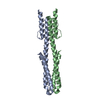
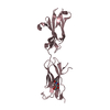
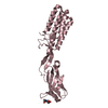
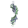
| Title | Structure of DraD invasin from uropathogenic Escherichia coli | ||||||
 Components Components | DraD invasin | ||||||
 Keywords Keywords | CELL INVASION /  HOMODIMER / BETA-SANDWICH / IMMUNOGLOBULIN-LIKE FOLD / SWAPPED C-TERMINAL STRANDS HOMODIMER / BETA-SANDWICH / IMMUNOGLOBULIN-LIKE FOLD / SWAPPED C-TERMINAL STRANDS | ||||||
| Function / homology |  Function and homology information Function and homology informationEnterobacteria AfaD invasin / Enterobacteria AfaD invasin protein / Dr adhesin / Dr-adhesin superfamily / Adhesion domain superfamily / Immunoglobulin-like / Sandwich / Mainly BetaSimilarity search - Domain/homology | ||||||
| Biological species |  Escherichia coli (E. coli) Escherichia coli (E. coli) | ||||||
| Method | X-RAY DIFFRACTION / SYNCHROTRON / SAD / Resolution: 1.05 Å | ||||||
 Authors Authors | Jedrzejczak, R. / Dauter, Z. / Dauter, M. / Piatek, R. / Zalewska, B. / Mroz, M. / Bury, K. / Nowicki, B. / Kur, J. | ||||||
 Citation Citation | Journal: Acta Crystallogr.,Sect.D / Year: 2006 Title: Structure of DraD invasin from uropathogenic Escherichia coli: a dimer with swapped beta-tails. Authors: Jedrzejczak, R. / Dauter, Z. / Dauter, M. / Piatek, R. / Zalewska, B. / Mroz, M. / Bury, K. / Nowicki, B. / Kur, J. | ||||||
| History |
|
- Structure visualization
| Structure viewer | Molecule: MolmilJmol/JSmol |
|---|
- Downloads & links
-Download
| PDBx/mmCIF format | 2axw.cif.gz | 167.2 KB | Display | PDBx/mmCIF format |
|---|---|---|---|---|
| PDB format | pdb2axw.ent.gz | 143.2 KB | Display | PDB format |
| PDBx/mmJSON format | 2axw.json.gz | Tree view | PDBx/mmJSON format | |
| Others |  Other downloads Other downloads |
-Validation report
| Arichive directory | https://data.pdbj.org/pub/pdb/validation_reports/ax/2axwftp://data.pdbj.org/pub/pdb/validation_reports/ax/2axw | HTTPS FTP |
|---|
-Related structure data
| Similar structure data |
|---|


-Links
 PDBj
PDBj- Assembly
| Deposited unit | 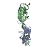
| ||||||||
|---|---|---|---|---|---|---|---|---|---|
| 1 |
| ||||||||
| Unit cell |
|
-Components
| #1: Protein | Mass: 14713.293 Da / Num. of mol.: 2 / Fragment: DraD invasin Source method: isolated from a genetically manipulated source Source: (gene. exp.) Escherichia coli (E. coli) / Gene: draD / Plasmid: pInvDsyg-C-His / Production host: Escherichia coli (E. coli) / Strain (production host): BL21 (DE3) RIL / References: UniProt: Q7BG36, UniProt: Q47038*PLUS#2: Chemical | ChemComp-CL / | Chloride  Mass: 35.453 Da / Num. of mol.: 1 / Source method: obtained synthetically / Formula: Cl Mass: 35.453 Da / Num. of mol.: 1 / Source method: obtained synthetically / Formula: Cl#3: Chemical | ChemComp-GOL / | Glycerol  Mass: 92.094 Da / Num. of mol.: 1 / Source method: obtained synthetically / Formula: C3H8O3 Mass: 92.094 Da / Num. of mol.: 1 / Source method: obtained synthetically / Formula: C3H8O3#4: Water | ChemComp-HOH / | Water Mass: 18.015 Da / Num. of mol.: 291 / Source method: isolated from a natural source / Formula: H2O Mass: 18.015 Da / Num. of mol.: 291 / Source method: isolated from a natural source / Formula: H2O |
|---|
-Experimental details
-Experiment
| Experiment | Method: X-RAY DIFFRACTION / Number of used crystals: 1 |
|---|
- Sample preparation
| Crystal | Density Matthews: 1.91 Å3/Da / Density % sol: 35 % |
|---|---|
| Crystal grow | Temperature: 293 K / Method: vapor diffusion, hanging drop / pH: 6 Details: NaCl, Hepes, glycerol, PEG-2000 MME, MES, pH 6.0, VAPOR DIFFUSION, HANGING DROP, temperature 293K |
-Data collection
| Diffraction | Mean temperature: 100 K |
|---|---|
| Diffraction source | Source: SYNCHROTRON / Site: APS  / Beamline: 22-ID / Wavelength: 1 Å / Beamline: 22-ID / Wavelength: 1 Å |
| Detector | Type: MARRESEARCH / Detector: CCD / Date: Jul 5, 2005 / Details: mirrors |
| Radiation | Monochromator: Si 111 CHANNEL CUT / Protocol: SINGLE WAVELENGTH / Monochromatic (M) / Laue (L): M / Scattering type: x-ray |
| Radiation wavelength | Wavelength: 1 Å / Relative weight: 1 |
| Reflection | Resolution: 1.05→30 Å / Num. all: 109502 / Num. obs: 108822 / % possible obs: 100 % / Observed criterion σ(F): 0 / Observed criterion σ(I): -3 / Redundancy: 5.6 % / Biso Wilson estimate: 9.1 Å2 / Rmerge(I) obs: 0.066 / Net I/σ(I): 21.4 |
| Reflection shell | Resolution: 1.05→1.09 Å / Redundancy: 4.6 % / Rmerge(I) obs: 0.388 / Mean I/σ(I) obs: 2.9 / Num. unique all: 10677 / % possible all: 97.6 |
- Processing
| Software |
| |||||||||||||||||||||||||||||||||||||||||||||||||||||||||||||||||||||||||||||||||||||||||||||||||||||||||||||||||||
|---|---|---|---|---|---|---|---|---|---|---|---|---|---|---|---|---|---|---|---|---|---|---|---|---|---|---|---|---|---|---|---|---|---|---|---|---|---|---|---|---|---|---|---|---|---|---|---|---|---|---|---|---|---|---|---|---|---|---|---|---|---|---|---|---|---|---|---|---|---|---|---|---|---|---|---|---|---|---|---|---|---|---|---|---|---|---|---|---|---|---|---|---|---|---|---|---|---|---|---|---|---|---|---|---|---|---|---|---|---|---|---|---|---|---|---|---|
| Refinement | Method to determine structure: SAD / Resolution: 1.05→29.49 Å / Cor.coef. Fo:Fc: 0.974 / Cor.coef. Fo:Fc free: 0.974 / SU B: 0.311 / SU ML: 0.016 / Cross valid method: THROUGHOUT / σ(F): 0 / σ(I): 0 / ESU R: 0.027 / ESU R Free: 0.027 / Stereochemistry target values: MAXIMUM LIKELIHOOD / Details: HYDROGENS HAVE BEEN ADDED IN THE RIDING POSITIONS
| |||||||||||||||||||||||||||||||||||||||||||||||||||||||||||||||||||||||||||||||||||||||||||||||||||||||||||||||||||
| Solvent computation | Ion probe radii: 0.8 Å / Shrinkage radii: 0.8 Å / VDW probe radii: 1.4 Å / Solvent model: BABINET MODEL WITH MASK | |||||||||||||||||||||||||||||||||||||||||||||||||||||||||||||||||||||||||||||||||||||||||||||||||||||||||||||||||||
| Displacement parameters | Biso mean: 12.984 Å2
| |||||||||||||||||||||||||||||||||||||||||||||||||||||||||||||||||||||||||||||||||||||||||||||||||||||||||||||||||||
| Refinement step | Cycle: LAST / Resolution: 1.05→29.49 Å
| |||||||||||||||||||||||||||||||||||||||||||||||||||||||||||||||||||||||||||||||||||||||||||||||||||||||||||||||||||
| Refine LS restraints |
| |||||||||||||||||||||||||||||||||||||||||||||||||||||||||||||||||||||||||||||||||||||||||||||||||||||||||||||||||||
| LS refinement shell | Resolution: 1.05→1.077 Å / Total num. of bins used: 20 /
|
