 Movie
Movie Controller


+ Open data

- Basic information
| Entry | Database: PDB / ID: 4bqb | ||||||
|---|---|---|---|---|---|---|---|
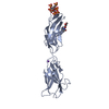
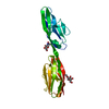
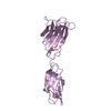
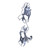
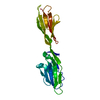
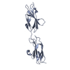
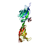
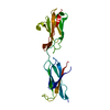
| Title | Crystal structure of the FN5 and FN6 domains of NEO1, form 2 | ||||||
 Components Components | NEOGENIN NEO1 NEO1 | ||||||
 Keywords Keywords | CELL ADHESION | ||||||
| Function / homology |  Function and homology information Function and homology informationnegative regulation of axon regeneration / co-receptor binding / BMP receptor binding / regulation of axon regeneration / myoblast fusion / plasma membrane protein complex / positive regulation of BMP signaling pathway / intracellular vesicle / protein secretion / negative regulation of protein secretion ...negative regulation of axon regeneration / co-receptor binding / BMP receptor binding / regulation of axon regeneration / myoblast fusion / plasma membrane protein complex / positive regulation of BMP signaling pathway / intracellular vesicle / protein secretion / negative regulation of protein secretion / axonal growth cone / axon guidance / neuron migration / multicellular organismal-level iron ion homeostasis / cell-cell adhesion / signaling receptor activity / growth cone / intracellular iron ion homeostasis / cadherin binding / neuronal cell body / regulation of DNA-templated transcription / Golgi apparatus / cell surface / nucleoplasm / membrane / plasma membraneSimilarity search - Function | ||||||
| Biological species |  MUS MUSCULUS (house mouse) MUS MUSCULUS (house mouse) | ||||||
| Method | X-RAY DIFFRACTION / SYNCHROTRON / MOLECULAR REPLACEMENT / Resolution: 2.7 Å | ||||||
 Authors Authors | Bell, C.H. / Healey, E. / van Erp, S. / Bishop, B. / Tang, C. / Gilbert, R.J.C. / Aricescu, A.R. / Pasterkamp, R.J. / Siebold, C. | ||||||
 Citation Citation | Journal: Science / Year: 2013 Title: Structure of the Repulsive Guidance Molecule (Rgm)-Neogenin Signaling Hub Authors: Bell, C.H. / Healey, E. / Van Erp, S. / Bishop, B. / Tang, C. / Gilbert, R.J.C. / Aricescu, A.R. / Pasterkamp, R.J. / Siebold, C. | ||||||
| History |
|
- Structure visualization
| Structure viewer | Molecule: MolmilJmol/JSmol |
|---|
- Downloads & links
-Download
| PDBx/mmCIF format | 4bqb.cif.gz | 329.6 KB | Display | PDBx/mmCIF format |
|---|---|---|---|---|
| PDB format | pdb4bqb.ent.gz | 274.6 KB | Display | PDB format |
| PDBx/mmJSON format | 4bqb.json.gz | Tree view | PDBx/mmJSON format | |
| Others |  Other downloads Other downloads |
-Validation report
| Arichive directory | https://data.pdbj.org/pub/pdb/validation_reports/bq/4bqbftp://data.pdbj.org/pub/pdb/validation_reports/bq/4bqb | HTTPS FTP |
|---|
-Related structure data


-Links
 PDBj
PDBj


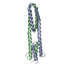
- Assembly
| Deposited unit | 
| ||||||||||||||||
|---|---|---|---|---|---|---|---|---|---|---|---|---|---|---|---|---|---|
| 1 | 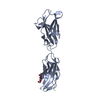
| ||||||||||||||||
| 2 | 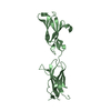
| ||||||||||||||||
| 3 | 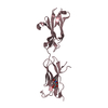
| ||||||||||||||||
| 4 | 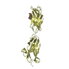
| ||||||||||||||||
| Unit cell |
| ||||||||||||||||
| Noncrystallographic symmetry (NCS) | NCS oper:
|
-Components
| #1: Protein | NEO1 Mass: 29221.842 Da / Num. of mol.: 4 / Fragment: FN-TYPE III DOMAINS 5 AND 6, RESIDUES 883-1133 Source method: isolated from a genetically manipulated source Details: N-LINKED GLYCOSYLATION AT N940 / Source: (gene. exp.) MUS MUSCULUS (house mouse) / Plasmid: PHLSEC / Cell line (production host): HEK293T / Production host:  HOMO SAPIENS (human) / References: UniProt: P97798 HOMO SAPIENS (human) / References: UniProt: P97798#2: Sugar | ChemComp-NAG / N-Acetylglucosamine  Type: D-saccharide, beta linking / Mass: 221.208 Da / Num. of mol.: 4 Type: D-saccharide, beta linking / Mass: 221.208 Da / Num. of mol.: 4Source method: isolated from a genetically manipulated source Formula: C8H15NO6 Nonpolymer details | N-ACETYL-D-GLUCOSAMIN | |
|---|
-Experimental details
-Experiment
| Experiment | Method: X-RAY DIFFRACTION / Number of used crystals: 1 |
|---|
- Sample preparation
| Crystal | Density Matthews: 2.15 Å3/Da / Density % sol: 56 % / Description: NONE |
|---|---|
| Crystal grow | pH: 8.5 / Details: 0.13 M POTASSIUM NITRATE, 13% PEG3350, pH 8.5 |
-Data collection
| Diffraction | Mean temperature: 100 K |
|---|---|
| Diffraction source | Source: SYNCHROTRON / Site: Diamond  / Beamline: I03 / Wavelength: 0.97922 / Beamline: I03 / Wavelength: 0.97922 |
| Detector | Type: ADSC CCD / Detector: CCD |
| Radiation | Protocol: SINGLE WAVELENGTH / Monochromatic (M) / Laue (L): M / Scattering type: x-ray |
| Radiation wavelength | Wavelength: 0.97922 Å / Relative weight: 1 |
| Reflection | Resolution: 2.7→30 Å / Num. obs: 26660 / % possible obs: 97.7 % / Observed criterion σ(I): 0 / Redundancy: 2.3 % / Biso Wilson estimate: 82.22 Å2 / Rmerge(I) obs: 0.08 / Net I/σ(I): 11.3 |
| Reflection shell | Resolution: 2.7→2.8 Å / Redundancy: 2.2 % / Rmerge(I) obs: 0.83 / Mean I/σ(I) obs: 1.2 / % possible all: 92.7 |
- Processing
| Software |
| |||||||||||||||||||||||||||||||||||||||||||||||||||||||||||||||||||||||||||||||||||||||||||||||||||||||||||||||||||||||||||||||||||||||||||||||||||||||||||||||||||||||||||||||||||||||||||||||||||||||||||||||||||||||||||||||||
|---|---|---|---|---|---|---|---|---|---|---|---|---|---|---|---|---|---|---|---|---|---|---|---|---|---|---|---|---|---|---|---|---|---|---|---|---|---|---|---|---|---|---|---|---|---|---|---|---|---|---|---|---|---|---|---|---|---|---|---|---|---|---|---|---|---|---|---|---|---|---|---|---|---|---|---|---|---|---|---|---|---|---|---|---|---|---|---|---|---|---|---|---|---|---|---|---|---|---|---|---|---|---|---|---|---|---|---|---|---|---|---|---|---|---|---|---|---|---|---|---|---|---|---|---|---|---|---|---|---|---|---|---|---|---|---|---|---|---|---|---|---|---|---|---|---|---|---|---|---|---|---|---|---|---|---|---|---|---|---|---|---|---|---|---|---|---|---|---|---|---|---|---|---|---|---|---|---|---|---|---|---|---|---|---|---|---|---|---|---|---|---|---|---|---|---|---|---|---|---|---|---|---|---|---|---|---|---|---|---|---|---|---|---|---|---|---|---|---|---|---|---|---|---|---|---|---|
| Refinement | Method to determine structure: MOLECULAR REPLACEMENT / Resolution: 2.7→30 Å / Cor.coef. Fo:Fc: 0.9261 / Cor.coef. Fo:Fc free: 0.9097 / SU R Cruickshank DPI: 1.208 / Cross valid method: THROUGHOUT / σ(F): 0 / SU R Blow DPI: 0.816 / SU Rfree Blow DPI: 0.283 / SU Rfree Cruickshank DPI: 0.296
| |||||||||||||||||||||||||||||||||||||||||||||||||||||||||||||||||||||||||||||||||||||||||||||||||||||||||||||||||||||||||||||||||||||||||||||||||||||||||||||||||||||||||||||||||||||||||||||||||||||||||||||||||||||||||||||||||
| Displacement parameters | Biso mean: 89.53 Å2
| |||||||||||||||||||||||||||||||||||||||||||||||||||||||||||||||||||||||||||||||||||||||||||||||||||||||||||||||||||||||||||||||||||||||||||||||||||||||||||||||||||||||||||||||||||||||||||||||||||||||||||||||||||||||||||||||||
| Refine analyze | Luzzati coordinate error obs: 0.468 Å | |||||||||||||||||||||||||||||||||||||||||||||||||||||||||||||||||||||||||||||||||||||||||||||||||||||||||||||||||||||||||||||||||||||||||||||||||||||||||||||||||||||||||||||||||||||||||||||||||||||||||||||||||||||||||||||||||
| Refinement step | Cycle: LAST / Resolution: 2.7→30 Å
| |||||||||||||||||||||||||||||||||||||||||||||||||||||||||||||||||||||||||||||||||||||||||||||||||||||||||||||||||||||||||||||||||||||||||||||||||||||||||||||||||||||||||||||||||||||||||||||||||||||||||||||||||||||||||||||||||
| Refine LS restraints |
| |||||||||||||||||||||||||||||||||||||||||||||||||||||||||||||||||||||||||||||||||||||||||||||||||||||||||||||||||||||||||||||||||||||||||||||||||||||||||||||||||||||||||||||||||||||||||||||||||||||||||||||||||||||||||||||||||
| LS refinement shell | Resolution: 2.7→2.81 Å / Total num. of bins used: 13
| |||||||||||||||||||||||||||||||||||||||||||||||||||||||||||||||||||||||||||||||||||||||||||||||||||||||||||||||||||||||||||||||||||||||||||||||||||||||||||||||||||||||||||||||||||||||||||||||||||||||||||||||||||||||||||||||||
| Refinement TLS params. | Method: refined / Refine-ID: X-RAY DIFFRACTION
| |||||||||||||||||||||||||||||||||||||||||||||||||||||||||||||||||||||||||||||||||||||||||||||||||||||||||||||||||||||||||||||||||||||||||||||||||||||||||||||||||||||||||||||||||||||||||||||||||||||||||||||||||||||||||||||||||
| Refinement TLS group |
|
