 Movie
Movie Controller

[English] 日本語
 Yorodumi
- PDB-6tpv: Crystal structures of FNIII domain one and two of the human leuco... -
+ Open data

- Basic information
| Entry | Database: PDB / ID: 6tpv | |||||||||
|---|---|---|---|---|---|---|---|---|---|---|
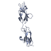
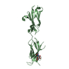
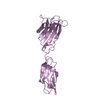
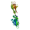
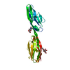
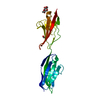
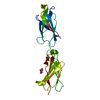
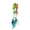
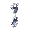
| Title | Crystal structures of FNIII domain one and two of the human leucocyte common antigen-related protein, LAR | |||||||||
 Components Components | Receptor-type tyrosine-protein phosphatase F | |||||||||
 Keywords Keywords |  CELL ADHESION / Fibronectin type-III / adhesion protein CELL ADHESION / Fibronectin type-III / adhesion protein | |||||||||
| Function / homology |  Function and homology informationchondroitin sulfate proteoglycan binding / cell surface receptor protein tyrosine phosphatase signaling pathway / neuron projection regeneration / Receptor-type tyrosine-protein phosphatases / synaptic membrane adhesion / transmembrane receptor protein tyrosine phosphatase activity / Synaptic adhesion-like molecules / regulation of axon regeneration / peptidyl-tyrosine dephosphorylation / Insulin receptor recycling ...chondroitin sulfate proteoglycan binding / cell surface receptor protein tyrosine phosphatase signaling pathway / neuron projection regeneration / Receptor-type tyrosine-protein phosphatases / synaptic membrane adhesion / transmembrane receptor protein tyrosine phosphatase activity / Synaptic adhesion-like molecules / regulation of axon regeneration / peptidyl-tyrosine dephosphorylation / Insulin receptor recycling / cell adhesion molecule binding / protein-tyrosine-phosphatase / negative regulation of receptor binding / protein tyrosine phosphatase activity / cell migration / heparin binding / cell adhesion / neuron projection / neuronal cell body / protein-containing complex binding / extracellular exosome / plasma membrane Function and homology informationchondroitin sulfate proteoglycan binding / cell surface receptor protein tyrosine phosphatase signaling pathway / neuron projection regeneration / Receptor-type tyrosine-protein phosphatases / synaptic membrane adhesion / transmembrane receptor protein tyrosine phosphatase activity / Synaptic adhesion-like molecules / regulation of axon regeneration / peptidyl-tyrosine dephosphorylation / Insulin receptor recycling ...chondroitin sulfate proteoglycan binding / cell surface receptor protein tyrosine phosphatase signaling pathway / neuron projection regeneration / Receptor-type tyrosine-protein phosphatases / synaptic membrane adhesion / transmembrane receptor protein tyrosine phosphatase activity / Synaptic adhesion-like molecules / regulation of axon regeneration / peptidyl-tyrosine dephosphorylation / Insulin receptor recycling / cell adhesion molecule binding / protein-tyrosine-phosphatase / negative regulation of receptor binding / protein tyrosine phosphatase activity / cell migration / heparin binding / cell adhesion / neuron projection / neuronal cell body / protein-containing complex binding / extracellular exosome / plasma membraneSimilarity search - Function | |||||||||
| Biological species |  Homo sapiens (human) Homo sapiens (human) | |||||||||
| Method | X-RAY DIFFRACTION / SYNCHROTRON / MOLECULAR REPLACEMENT / Resolution: 1.8 Å | |||||||||
 Authors Authors | Vilstrup, J.P. / Thirup, S.S. / Simonsen, A. / Birkefeldt, T. / Strandbygaard, D. | |||||||||
| Funding support |  Denmark, 2items Denmark, 2items
| |||||||||
 Citation Citation | Journal: Acta Crystallogr D Struct Biol / Year: 2020 Title: Crystal and solution structures of fragments of the human leucocyte common antigen-related protein. Authors: Vilstrup, J. / Simonsen, A. / Birkefeldt, T. / Strandbygard, D. / Lyngso, J. / Pedersen, J.S. / Thirup, S. | |||||||||
| History |
|
- Structure visualization
| Structure viewer | Molecule: MolmilJmol/JSmol |
|---|
- Downloads & links
-Download
| PDBx/mmCIF format | 6tpv.cif.gz | 175 KB | Display | PDBx/mmCIF format |
|---|---|---|---|---|
| PDB format | pdb6tpv.ent.gz | 132.3 KB | Display | PDB format |
| PDBx/mmJSON format | 6tpv.json.gz | Tree view | PDBx/mmJSON format | |
| Others |  Other downloads Other downloads |
-Validation report
| Arichive directory | https://data.pdbj.org/pub/pdb/validation_reports/tp/6tpvftp://data.pdbj.org/pub/pdb/validation_reports/tp/6tpv | HTTPS FTP |
|---|
-Related structure data
| Related structure data | 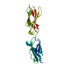 6tptC  6tpuC 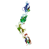 6tpwC 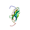 2djuS S: Starting model for refinement C: citing same article ( |
|---|---|
| Similar structure data |


-Links
 PDBj
PDBj


- Assembly
| Deposited unit | 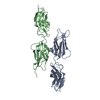
| ||||||||||||
|---|---|---|---|---|---|---|---|---|---|---|---|---|---|
| 1 | 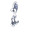
| ||||||||||||
| 2 | 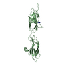
| ||||||||||||
| Unit cell |
|
-Components
| #1: Protein | Mass: 22729.354 Da / Num. of mol.: 2 Source method: isolated from a genetically manipulated source Source: (gene. exp.) Homo sapiens (human) / Gene: PTPRF, LAR / Production host:  Escherichia coli BL21(DE3) (bacteria) / References: UniProt: P10586, protein-tyrosine-phosphatase Escherichia coli BL21(DE3) (bacteria) / References: UniProt: P10586, protein-tyrosine-phosphatase#2: Chemical | ChemComp-IMD / | Imidazole  Mass: 69.085 Da / Num. of mol.: 1 / Source method: obtained synthetically / Formula: C3H5N2 Mass: 69.085 Da / Num. of mol.: 1 / Source method: obtained synthetically / Formula: C3H5N2#3: Water | ChemComp-HOH / | Water Mass: 18.015 Da / Num. of mol.: 340 / Source method: isolated from a natural source / Formula: H2O Mass: 18.015 Da / Num. of mol.: 340 / Source method: isolated from a natural source / Formula: H2OHas ligand of interest | N | |
|---|
-Experimental details
-Experiment
| Experiment | Method: X-RAY DIFFRACTION / Number of used crystals: 1 |
|---|
- Sample preparation
| Crystal | Density Matthews: 2.38 Å3/Da / Density % sol: 48.24 % |
|---|---|
| Crystal grow | Temperature: 277 K / Method: vapor diffusion, sitting drop / pH: 8.3 Details: 0.2 M Sodium citrate tribasic dihydrate pH 8.3, 20 % PEG 3,350 |
-Data collection
| Diffraction | Mean temperature: 100 K / Serial crystal experiment: N |
|---|---|
| Diffraction source | Source: SYNCHROTRON / Site: PETRA III, EMBL c/o DESY  / Beamline: P14 (MX2) / Wavelength: 0.9794 Å / Beamline: P14 (MX2) / Wavelength: 0.9794 Å |
| Detector | Type: DECTRIS EIGER X 16M / Detector: PIXEL / Date: Sep 22, 2018 |
| Radiation | Protocol: SINGLE WAVELENGTH / Monochromatic (M) / Laue (L): M / Scattering type: x-ray |
| Radiation wavelength | Wavelength: 0.9794 Å / Relative weight: 1 |
| Reflection | Resolution: 1.8→40.05 Å / Num. obs: 37701 / % possible obs: 97.8 % / Redundancy: 6.8 % / CC1/2: 0.999 / Net I/σ(I): 17.3 |
| Reflection shell | Resolution: 1.8→1.86 Å / Redundancy: 7.1 % / Mean I/σ(I) obs: 1.81 / Num. unique obs: 2761 / CC1/2: 0.888 / Rrim(I) all: 0.98 / % possible all: 97.4 |
- Processing
| Software |
| ||||||||||||||||||||||||
|---|---|---|---|---|---|---|---|---|---|---|---|---|---|---|---|---|---|---|---|---|---|---|---|---|---|
| Refinement | Method to determine structure: MOLECULAR REPLACEMENT Starting model: 2DJU Resolution: 1.8→40.05 Å / Cross valid method: FREE R-VALUE
| ||||||||||||||||||||||||
| Displacement parameters | Biso mean: 41.93 Å2 | ||||||||||||||||||||||||
| Refinement step | Cycle: LAST / Resolution: 1.8→40.05 Å
| ||||||||||||||||||||||||
| Refine LS restraints |
|
