 Movie
Movie Controller


+ Open data

- Basic information
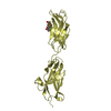
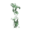
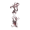
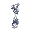
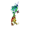
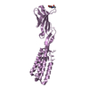
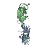
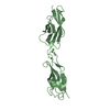
| Entry | Database: PDB / ID: 3p4l | ||||||
|---|---|---|---|---|---|---|---|
| Title | Crystal structure of a hemojuvelin-binding fragment of neogenin | ||||||
 Components Components | Neogenin NEO1 NEO1 | ||||||
 Keywords Keywords | CELL ADHESION / IRON HOMEOSTASIS / HEMOJUVELIN RECEPTOR / FNIII domain / fibronectin type III | ||||||
| Function / homology |  Function and homology information Function and homology informationnegative regulation of axon regeneration / Netrin-1 signaling / co-receptor binding / BMP receptor binding / myoblast fusion / plasma membrane protein complex / positive regulation of BMP signaling pathway / intracellular vesicle / Myogenesis / protein secretion ...negative regulation of axon regeneration / Netrin-1 signaling / co-receptor binding / BMP receptor binding / myoblast fusion / plasma membrane protein complex / positive regulation of BMP signaling pathway / intracellular vesicle / Myogenesis / protein secretion / negative regulation of protein secretion / axonal growth cone / axon guidance / neuron migration / multicellular organismal-level iron ion homeostasis / cell-cell adhesion / signaling receptor activity / intracellular iron ion homeostasis / cell adhesion / cadherin binding / regulation of DNA-templated transcription / Golgi apparatus / cell surface / nucleoplasm / plasma membraneSimilarity search - Function | ||||||
| Biological species |  Homo sapiens (human) Homo sapiens (human) | ||||||
| Method | X-RAY DIFFRACTION / MOLECULAR REPLACEMENT / Resolution: 1.8 Å | ||||||
 Authors Authors | Yang, F. / West Jr., A.P. / Bjorkman, P.J. | ||||||
 Citation Citation | Journal: J.Struct.Biol. / Year: 2011 Title: Crystal structure of a hemojuvelin-binding fragment of neogenin at 1.8A. Authors: Yang, F. / West, A.P. / Bjorkman, P.J. | ||||||
| History |
|
- Structure visualization
| Structure viewer | Molecule: MolmilJmol/JSmol |
|---|
- Downloads & links
-Download
| PDBx/mmCIF format | 3p4l.cif.gz | 59.4 KB | Display | PDBx/mmCIF format |
|---|---|---|---|---|
| PDB format | pdb3p4l.ent.gz | 42.5 KB | Display | PDB format |
| PDBx/mmJSON format | 3p4l.json.gz | Tree view | PDBx/mmJSON format | |
| Others |  Other downloads Other downloads |
-Validation report
| Arichive directory | https://data.pdbj.org/pub/pdb/validation_reports/p4/3p4lftp://data.pdbj.org/pub/pdb/validation_reports/p4/3p4l | HTTPS FTP |
|---|
-Related structure data


-Links
 PDBj
PDBj


- Assembly
| Deposited unit | 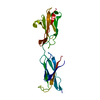
| ||||||||||||
|---|---|---|---|---|---|---|---|---|---|---|---|---|---|
| 1 |
| ||||||||||||
| Unit cell |
| ||||||||||||
| Components on special symmetry positions |
|
-Components
| #1: Protein | NEO1 / Immunoglobulin superfamily DCC subclass member 2 Mass: 23902.195 Da / Num. of mol.: 1 / Fragment: BINDING-DOMAIN, RESIDUES 853-1051 Source method: isolated from a genetically manipulated source Source: (gene. exp.) Homo sapiens (human) / Gene: NEO1, IGDCC2, NGN / Plasmid: PACGP67 / Cell line (production host): BTI-TN-5B1-4 / Production host:  TRICHOPLUSIA NI (cabbage looper) / References: UniProt: Q92859 TRICHOPLUSIA NI (cabbage looper) / References: UniProt: Q92859 |
|---|---|
| #2: Water | ChemComp-HOH / Water Mass: 18.015 Da / Num. of mol.: 293 / Source method: isolated from a natural source / Formula: H2O Mass: 18.015 Da / Num. of mol.: 293 / Source method: isolated from a natural source / Formula: H2O |
-Experimental details
-Experiment
| Experiment | Method: X-RAY DIFFRACTION / Number of used crystals: 1 |
|---|
- Sample preparation
| Crystal | Density Matthews: 2.5 Å3/Da / Density % sol: 51 % / Description: NONE |
|---|---|
| Crystal grow | Temperature: 293 K / pH: 8.5 Details: 25% PEG-3350, 0.2m ammonium sulfate, 0.1m TRIS, pH 8.5, temperature 293k, VAPOR DIFFUSION, HANGING DROP |
-Data collection
| Diffraction | Mean temperature: 100 K |
|---|---|
| Diffraction source | Source: ROTATING ANODE / Type: RIGAKU / Wavelength: 1.54 |
| Detector | Type: RIGAKU / Detector: IMAGE PLATE / Date: Feb 26, 2008 |
| Radiation | Protocol: SINGLE WAVELENGTH / Monochromatic (M) / Laue (L): M / Scattering type: x-ray |
| Radiation wavelength | Wavelength: 1.54 Å / Relative weight: 1 |
| Reflection | Resolution: 1.8→40 Å / Num. obs: 22544 / % possible obs: 99.3 % / Redundancy: 3.8 % / Biso Wilson estimate: 17.8 Å2 / Rmerge(I) obs: 0.059 / Net I/σ(I): 28.2 |
| Reflection shell | Resolution: 1.8→1.86 Å / Redundancy: 3.7 % / Rmerge(I) obs: 0.38 / Mean I/σ(I) obs: 4.2 / % possible all: 98.2 |
- Processing
| Software |
| ||||||||||||||||||||||||||||||||||||||||||||||||||||||||||||
|---|---|---|---|---|---|---|---|---|---|---|---|---|---|---|---|---|---|---|---|---|---|---|---|---|---|---|---|---|---|---|---|---|---|---|---|---|---|---|---|---|---|---|---|---|---|---|---|---|---|---|---|---|---|---|---|---|---|---|---|---|---|
| Refinement | Method to determine structure: MOLECULAR REPLACEMENT Starting model: PDB ENTRY 1X5J and PDB ENTRY 1X5K Resolution: 1.8→32.87 Å / Rfactor Rfree error: 0.007 / Data cutoff high absF: 1503078.09 / Data cutoff low absF: 0 / Isotropic thermal model: RESTRAINED / Cross valid method: THROUGHOUT / σ(F): 0 / Details: BULK SOLVENT MODEL USED
| ||||||||||||||||||||||||||||||||||||||||||||||||||||||||||||
| Solvent computation | Solvent model: FLAT MODEL / Bsol: 50.8098 Å2 / ksol: 0.35 e/Å3 | ||||||||||||||||||||||||||||||||||||||||||||||||||||||||||||
| Displacement parameters | Biso mean: 25.7 Å2
| ||||||||||||||||||||||||||||||||||||||||||||||||||||||||||||
| Refine analyze |
| ||||||||||||||||||||||||||||||||||||||||||||||||||||||||||||
| Refinement step | Cycle: LAST / Resolution: 1.8→32.87 Å
| ||||||||||||||||||||||||||||||||||||||||||||||||||||||||||||
| Refine LS restraints |
| ||||||||||||||||||||||||||||||||||||||||||||||||||||||||||||
| Refine LS restraints NCS | NCS model details: NONE | ||||||||||||||||||||||||||||||||||||||||||||||||||||||||||||
| LS refinement shell | Resolution: 1.8→1.91 Å / Rfactor Rfree error: 0.022 / Total num. of bins used: 6
|
