 Movie
Movie Controller


+ Open data

- Basic information
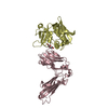
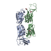
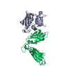
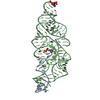
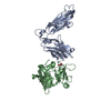
| Entry | Database: PDB / ID: 6ulm | ||||||
|---|---|---|---|---|---|---|---|
| Title | Crystal structure of human cadherin 17 EC1-2 | ||||||
 Components Components | Cadherin-17 | ||||||
 Keywords Keywords | CELL ADHESION / cdh17 / intestinal epithelia / cancer / calcium-binding | ||||||
| Function / homology |  Function and homology information Function and homology informationoligopeptide transmembrane transport / positive regulation of integrin activation by cell surface receptor linked signal transduction / proton-dependent oligopeptide secondary active transmembrane transporter activity / marginal zone B cell differentiation / germinal center B cell differentiation / oligopeptide transport / cell-cell adhesion mediated by cadherin / calcium-dependent cell-cell adhesion via plasma membrane cell adhesion molecules / Adherens junctions interactions / catenin complex ...oligopeptide transmembrane transport / positive regulation of integrin activation by cell surface receptor linked signal transduction / proton-dependent oligopeptide secondary active transmembrane transporter activity / marginal zone B cell differentiation / germinal center B cell differentiation / oligopeptide transport / cell-cell adhesion mediated by cadherin / calcium-dependent cell-cell adhesion via plasma membrane cell adhesion molecules / Adherens junctions interactions / catenin complex / cell-cell junction assembly / adherens junction organization / homophilic cell adhesion via plasma membrane adhesion molecules / spleen development / integrin-mediated signaling pathway / adherens junction / cell morphogenesis / integrin binding / cell junction / basolateral plasma membrane / cell adhesion / cadherin binding / calcium ion binding / cell surface / nucleoplasm / plasma membraneSimilarity search - Function | ||||||
| Biological species |  Homo sapiens (human) Homo sapiens (human) | ||||||
| Method | X-RAY DIFFRACTION / SYNCHROTRON / MOLECULAR REPLACEMENT / Resolution: 2.15 Å | ||||||
 Authors Authors | Gray, M.E. / Sotomayor, M. | ||||||
 Citation Citation | Journal: Acta Crystallogr.,Sect.F / Year: 2021 Title: Crystal structure of the nonclassical cadherin-17 N-terminus and implications for its adhesive binding mechanism. Authors: Gray, M.E. / Sotomayor, M. | ||||||
| History |
|
- Structure visualization
| Structure viewer | Molecule: MolmilJmol/JSmol |
|---|
- Downloads & links
-Download
| PDBx/mmCIF format | 6ulm.cif.gz | 178.3 KB | Display | PDBx/mmCIF format |
|---|---|---|---|---|
| PDB format | pdb6ulm.ent.gz | 140.1 KB | Display | PDB format |
| PDBx/mmJSON format | 6ulm.json.gz | Tree view | PDBx/mmJSON format | |
| Others |  Other downloads Other downloads |
-Validation report
| Arichive directory | https://data.pdbj.org/pub/pdb/validation_reports/ul/6ulmftp://data.pdbj.org/pub/pdb/validation_reports/ul/6ulm | HTTPS FTP |
|---|
-Related structure data
| Related structure data | 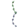 3q2vS S: Starting model for refinement |
|---|---|
| Similar structure data |


-Links
 PDBj
PDBj


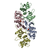
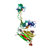
- Assembly
| Deposited unit | 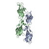
| |||||||||||||||||||||||||||||||||||||||
|---|---|---|---|---|---|---|---|---|---|---|---|---|---|---|---|---|---|---|---|---|---|---|---|---|---|---|---|---|---|---|---|---|---|---|---|---|---|---|---|---|
| 1 |
| |||||||||||||||||||||||||||||||||||||||
| 2 | 
| |||||||||||||||||||||||||||||||||||||||
| 3 | 
| |||||||||||||||||||||||||||||||||||||||
| Unit cell |
| |||||||||||||||||||||||||||||||||||||||
| Noncrystallographic symmetry (NCS) | NCS domain:
NCS domain segments:
NCS oper:
|
-Components
| #1: Protein | / Intestinal peptide-associated transporter HPT-1 / Liver-intestine cadherin / LI-cadherin Mass: 25690.705 Da / Num. of mol.: 2 Source method: isolated from a genetically manipulated source Source: (gene. exp.) Homo sapiens (human) / Gene: CDH17 / Variant: VAR_055567 / Plasmid: pET21a+ / Production host:  Escherichia coli BL21(DE3) (bacteria) / Strain (production host): BL21(DE3) / Variant (production host): Rosetta2 / References: UniProt: Q12864 Escherichia coli BL21(DE3) (bacteria) / Strain (production host): BL21(DE3) / Variant (production host): Rosetta2 / References: UniProt: Q12864#2: Chemical | ChemComp-CA /   Mass: 40.078 Da / Num. of mol.: 6 / Source method: obtained synthetically / Formula: Ca Mass: 40.078 Da / Num. of mol.: 6 / Source method: obtained synthetically / Formula: Ca#3: Water | ChemComp-HOH / | Water Mass: 18.015 Da / Num. of mol.: 203 / Source method: isolated from a natural source / Formula: H2O Mass: 18.015 Da / Num. of mol.: 203 / Source method: isolated from a natural source / Formula: H2OHas ligand of interest | N | |
|---|
-Experimental details
-Experiment
| Experiment | Method: X-RAY DIFFRACTION / Number of used crystals: 1 |
|---|
- Sample preparation
| Crystal | Density Matthews: 2.56 Å3/Da / Density % sol: 52 % |
|---|---|
| Crystal grow | Temperature: 277 K / Method: vapor diffusion, sitting drop / pH: 7 / Details: 0.1 M HEPES pH 7, 0.1 M KCl, 15% PEG 5000 MME |
-Data collection
| Diffraction | Mean temperature: 100 K / Serial crystal experiment: N |
|---|---|
| Diffraction source | Source: SYNCHROTRON / Site: APS  / Beamline: 24-ID-E / Wavelength: 0.97919 Å / Beamline: 24-ID-E / Wavelength: 0.97919 Å |
| Detector | Type: ADSC QUANTUM 315 / Detector: CCD / Date: Dec 2, 2015 |
| Radiation | Protocol: SINGLE WAVELENGTH / Monochromatic (M) / Laue (L): M / Scattering type: x-ray |
| Radiation wavelength | Wavelength: 0.97919 Å / Relative weight: 1 |
| Reflection | Resolution: 2.15→50 Å / Num. obs: 28521 / % possible obs: 99.5 % / Redundancy: 3.6 % / Rmerge(I) obs: 0.132 / Net I/σ(I): 10.46 |
| Reflection shell | Resolution: 2.15→2.19 Å / Redundancy: 3.5 % / Rmerge(I) obs: 0.73 / Mean I/σ(I) obs: 2.05 / Num. unique obs: 1406 / % possible all: 99.9 |
- Processing
| Software |
| |||||||||||||||||||||||||||||||||||||||||||||||||||||||||||||||||||||||||||||||||||||||||||||||||||||||||||||||||||||||||||||
|---|---|---|---|---|---|---|---|---|---|---|---|---|---|---|---|---|---|---|---|---|---|---|---|---|---|---|---|---|---|---|---|---|---|---|---|---|---|---|---|---|---|---|---|---|---|---|---|---|---|---|---|---|---|---|---|---|---|---|---|---|---|---|---|---|---|---|---|---|---|---|---|---|---|---|---|---|---|---|---|---|---|---|---|---|---|---|---|---|---|---|---|---|---|---|---|---|---|---|---|---|---|---|---|---|---|---|---|---|---|---|---|---|---|---|---|---|---|---|---|---|---|---|---|---|---|---|
| Refinement | Method to determine structure: MOLECULAR REPLACEMENT Starting model: 3Q2V Resolution: 2.15→47.53 Å / Cor.coef. Fo:Fc: 0.935 / Cor.coef. Fo:Fc free: 0.897 / SU B: 10.68 / SU ML: 0.142 / Cross valid method: THROUGHOUT / σ(F): 0 / ESU R: 0.228 / ESU R Free: 0.2 Details: HYDROGENS HAVE BEEN ADDED IN THE RIDING POSITIONS U VALUES : WITH TLS ADDED
| |||||||||||||||||||||||||||||||||||||||||||||||||||||||||||||||||||||||||||||||||||||||||||||||||||||||||||||||||||||||||||||
| Solvent computation | Ion probe radii: 0.8 Å / Shrinkage radii: 0.8 Å / VDW probe radii: 1.2 Å | |||||||||||||||||||||||||||||||||||||||||||||||||||||||||||||||||||||||||||||||||||||||||||||||||||||||||||||||||||||||||||||
| Displacement parameters | Biso max: 104.63 Å2 / Biso mean: 30.462 Å2 / Biso min: 13.07 Å2
| |||||||||||||||||||||||||||||||||||||||||||||||||||||||||||||||||||||||||||||||||||||||||||||||||||||||||||||||||||||||||||||
| Refinement step | Cycle: final / Resolution: 2.15→47.53 Å
| |||||||||||||||||||||||||||||||||||||||||||||||||||||||||||||||||||||||||||||||||||||||||||||||||||||||||||||||||||||||||||||
| Refine LS restraints |
| |||||||||||||||||||||||||||||||||||||||||||||||||||||||||||||||||||||||||||||||||||||||||||||||||||||||||||||||||||||||||||||
| Refine LS restraints NCS | Number: 1534 / Type: TIGHT THERMAL / Rms dev position: 4.84 Å / Weight position: 0.5 | |||||||||||||||||||||||||||||||||||||||||||||||||||||||||||||||||||||||||||||||||||||||||||||||||||||||||||||||||||||||||||||
| LS refinement shell | Resolution: 2.15→2.205 Å / Rfactor Rfree error: 0 / Total num. of bins used: 20
| |||||||||||||||||||||||||||||||||||||||||||||||||||||||||||||||||||||||||||||||||||||||||||||||||||||||||||||||||||||||||||||
| Refinement TLS params. | Method: refined / Refine-ID: X-RAY DIFFRACTION
| |||||||||||||||||||||||||||||||||||||||||||||||||||||||||||||||||||||||||||||||||||||||||||||||||||||||||||||||||||||||||||||
| Refinement TLS group |
|
