 Movie
Movie Controller


+ Open data

- Basic information
| Entry | Database: PDB / ID: 6mje | ||||||
|---|---|---|---|---|---|---|---|
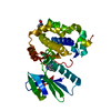
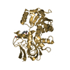
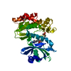
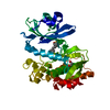
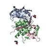
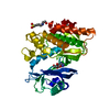
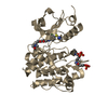
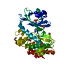
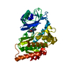
| Title | Structure of Candida glabrata Csm1: S. cerevisiae Dsn1 complex | ||||||
 Components Components |
| ||||||
 Keywords Keywords |  CELL CYCLE / monopolin / kinetochore CELL CYCLE / monopolin / kinetochore | ||||||
| Function / homology |  Function and homology information Function and homology informationMIS12/MIND type complex / monopolin complex / spindle attachment to meiosis I kinetochore / protein localization to nucleolar rDNA repeats / meiotic sister chromatid cohesion, centromeric / rDNA chromatin condensation / attachment of spindle microtubules to kinetochore / mitotic sister chromatid segregation / Neutrophil degranulation / chromosome segregation ...MIS12/MIND type complex / monopolin complex / spindle attachment to meiosis I kinetochore / protein localization to nucleolar rDNA repeats / meiotic sister chromatid cohesion, centromeric / rDNA chromatin condensation / attachment of spindle microtubules to kinetochore / mitotic sister chromatid segregation / Neutrophil degranulation / chromosome segregation / kinetochore / spindle pole / nuclear envelope / cell division / nucleolus / identical protein binding / nucleusSimilarity search - Function | ||||||
| Biological species |  Candida glabrata (fungus) Candida glabrata (fungus) Saccharomyces cerevisiae (brewer's yeast) Saccharomyces cerevisiae (brewer's yeast) | ||||||
| Method | X-RAY DIFFRACTION / SYNCHROTRON / MOLECULAR REPLACEMENT / Resolution: 2.5 Å | ||||||
 Authors Authors | Singh, N. / Corbett, K.D. | ||||||
| Funding support |  United States, 1items United States, 1items
| ||||||
 Citation Citation | Journal: Chromosoma / Year: 2019 Title: The molecular basis of monopolin recruitment to the kinetochore. Authors: Plowman, R. / Singh, N. / Tromer, E.C. / Payan, A. / Duro, E. / Spanos, C. / Rappsilber, J. / Snel, B. / Kops, G.J.P.L. / Corbett, K.D. / Marston, A.L. | ||||||
| History |
|
- Structure visualization
| Structure viewer | Molecule: MolmilJmol/JSmol |
|---|
- Downloads & links
-Download
| PDBx/mmCIF format | 6mje.cif.gz | 240.4 KB | Display | PDBx/mmCIF format |
|---|---|---|---|---|
| PDB format | pdb6mje.ent.gz | 193.4 KB | Display | PDB format |
| PDBx/mmJSON format | 6mje.json.gz | Tree view | PDBx/mmJSON format | |
| Others |  Other downloads Other downloads |
-Validation report
| Arichive directory | https://data.pdbj.org/pub/pdb/validation_reports/mj/6mjeftp://data.pdbj.org/pub/pdb/validation_reports/mj/6mje | HTTPS FTP |
|---|
-Related structure data
| Related structure data | 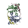 6mj8C 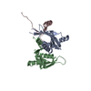 6mjbC 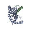 6mjcC 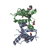 3n4rS S: Starting model for refinement C: citing same article ( |
|---|---|
| Similar structure data | |
| Experimental dataset #1 | Data reference: 10.15785/SBGRID/610 / Data set type: diffraction image data / Details: SBGrid |

-Links
 PDBj
PDBj


- Assembly
| Deposited unit | 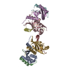
| ||||||||
|---|---|---|---|---|---|---|---|---|---|
| 1 | 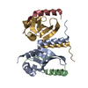
| ||||||||
| 2 | 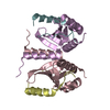
| ||||||||
| Unit cell |
|
-Components
| #1: Protein | Mass: 15571.603 Da / Num. of mol.: 4 / Fragment: UNP residues 69-181 Source method: isolated from a genetically manipulated source Source: (gene. exp.) Candida glabrata (fungus) / Gene: AO440_000897, AO440_004693 / Production host:  Escherichia coli (E. coli) / References: UniProt: A0A0W0CH22, UniProt: Q6FVN3*PLUS Escherichia coli (E. coli) / References: UniProt: A0A0W0CH22, UniProt: Q6FVN3*PLUS#2: Protein/peptide | Mass: 4763.462 Da / Num. of mol.: 4 / Fragment: UNP residues 71-110 Source method: isolated from a genetically manipulated source Source: (gene. exp.) Saccharomyces cerevisiae (brewer's yeast)Gene: DSN1 / Production host: Escherichia coli (E. coli) / References: UniProt: A0A1L4A9Z4, UniProt: P40568*PLUS |
|---|
-Experimental details
-Experiment
| Experiment | Method: X-RAY DIFFRACTION / Number of used crystals: 1 |
|---|
- Sample preparation
| Crystal | Density Matthews: 2.46 Å3/Da / Density % sol: 49.99 % |
|---|---|
| Crystal grow | Temperature: 293 K / Method: vapor diffusion, hanging drop / pH: 8.5 Details: 0.2 M magnesium chloride, 0.1 M Tris-HCl, pH 8.5, 25% PEG3350, cryoprotect with additional 20% PEG400 |
-Data collection
| Diffraction | Mean temperature: 100 K |
|---|---|
| Diffraction source | Source: SYNCHROTRON / Site: SSRL / Beamline: BL14-1 / Wavelength: 1.127 Å |
| Detector | Type: MARMOSAIC 325 mm CCD / Detector: CCD / Date: Apr 15, 2016 |
| Radiation | Monochromator: double crystal Si(111) / Protocol: SINGLE WAVELENGTH / Monochromatic (M) / Laue (L): M / Scattering type: x-ray |
| Radiation wavelength | Wavelength: 1.127 Å / Relative weight: 1 |
| Reflection | Resolution: 2.5→50 Å / Num. obs: 26122 / % possible obs: 96 % / Redundancy: 4.6 % / Rmerge(I) obs: 0.199 / Rpim(I) all: 0.091 / Rrim(I) all: 0.219 / Net I/σ(I): 18.2 |
| Reflection shell | Resolution: 2.5→2.56 Å / Rmerge(I) obs: 0.793 / Num. unique obs: 1446 / Rpim(I) all: 0.488 / Rrim(I) all: 0.938 |
- Processing
| Software |
| ||||||||||||||||||||||||||||||||||||||||||||||||||||||||||||||||||||||
|---|---|---|---|---|---|---|---|---|---|---|---|---|---|---|---|---|---|---|---|---|---|---|---|---|---|---|---|---|---|---|---|---|---|---|---|---|---|---|---|---|---|---|---|---|---|---|---|---|---|---|---|---|---|---|---|---|---|---|---|---|---|---|---|---|---|---|---|---|---|---|---|
| Refinement | Method to determine structure: MOLECULAR REPLACEMENT Starting model: PDB entry 3N4R Resolution: 2.5→43.351 Å / SU ML: 0.47 / Cross valid method: FREE R-VALUE / σ(F): 1.35 / Phase error: 43.07
| ||||||||||||||||||||||||||||||||||||||||||||||||||||||||||||||||||||||
| Solvent computation | Shrinkage radii: 0.9 Å / VDW probe radii: 1.11 Å | ||||||||||||||||||||||||||||||||||||||||||||||||||||||||||||||||||||||
| Refinement step | Cycle: LAST / Resolution: 2.5→43.351 Å
| ||||||||||||||||||||||||||||||||||||||||||||||||||||||||||||||||||||||
| Refine LS restraints |
| ||||||||||||||||||||||||||||||||||||||||||||||||||||||||||||||||||||||
| LS refinement shell |
| ||||||||||||||||||||||||||||||||||||||||||||||||||||||||||||||||||||||
| Refinement TLS params. | Method: refined / Origin x: 11.1476 Å / Origin y: -24.6765 Å / Origin z: 4.351 Å
| ||||||||||||||||||||||||||||||||||||||||||||||||||||||||||||||||||||||
| Refinement TLS group | Selection details: all |
