 Movie
Movie Controller


+ Open data

- Basic information
| Entry | Database: PDB / ID: 3n4r | ||||||
|---|---|---|---|---|---|---|---|
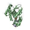
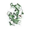
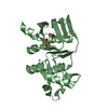
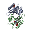
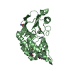
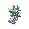
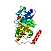
| Title | Structure of Csm1 C-terminal domain, R3 form | ||||||
 Components Components | Monopolin complex subunit CSM1 | ||||||
 Keywords Keywords | REPLICATION / meiosis / rDNA | ||||||
| Function / homology |  Function and homology informationmonopolin complex / spindle attachment to meiosis I kinetochore / protein localization to nucleolar rDNA repeats / meiotic chromosome segregation / meiotic sister chromatid cohesion, centromeric / rDNA chromatin condensation / homologous chromosome segregation / nuclear envelope / nucleolus / identical protein binding Function and homology informationmonopolin complex / spindle attachment to meiosis I kinetochore / protein localization to nucleolar rDNA repeats / meiotic chromosome segregation / meiotic sister chromatid cohesion, centromeric / rDNA chromatin condensation / homologous chromosome segregation / nuclear envelope / nucleolus / identical protein bindingSimilarity search - Function | ||||||
| Biological species |  Saccharomyces cerevisiae (brewer's yeast) Saccharomyces cerevisiae (brewer's yeast) | ||||||
| Method | X-RAY DIFFRACTION / SYNCHROTRON / SAD / Resolution: 2.602 Å | ||||||
 Authors Authors | Corbett, K.D. / Harrison, S.C. | ||||||
 Citation Citation | Journal: Cell(Cambridge,Mass.) / Year: 2010 Title: The Monopolin Complex Crosslinks Kinetochore Components to Regulate Chromosome-Microtubule Attachments. Authors: Corbett, K.D. / Yip, C.K. / Ee, L.S. / Walz, T. / Amon, A. / Harrison, S.C. | ||||||
| History |
|
- Structure visualization
| Structure viewer | Molecule: MolmilJmol/JSmol |
|---|
- Downloads & links
-Download
| PDBx/mmCIF format | 3n4r.cif.gz | 167.9 KB | Display | PDBx/mmCIF format |
|---|---|---|---|---|
| PDB format | pdb3n4r.ent.gz | 141.9 KB | Display | PDB format |
| PDBx/mmJSON format | 3n4r.json.gz | Tree view | PDBx/mmJSON format | |
| Others |  Other downloads Other downloads |
-Validation report
| Arichive directory | https://data.pdbj.org/pub/pdb/validation_reports/n4/3n4rftp://data.pdbj.org/pub/pdb/validation_reports/n4/3n4r | HTTPS FTP |
|---|
-Related structure data


-Links
 PDBj
PDBj- Assembly
| Deposited unit | 
| ||||||||
|---|---|---|---|---|---|---|---|---|---|
| 1 | 
| ||||||||
| 2 | 
| ||||||||
| Unit cell |
|
-Components
| #1: Protein | / Chromosome segregation in meiosis protein 1 Mass: 13375.534 Da / Num. of mol.: 4 / Fragment: C-terminal domain (residues 69-181) / Mutation: L157M Source method: isolated from a genetically manipulated source Source: (gene. exp.) Saccharomyces cerevisiae (brewer's yeast)Gene: CSM1, SPO86, YCR086W, YCR86W / Plasmid: pET3a / Production host:  Escherichia coli (E. coli) / Strain (production host): Rosetta 2 pLysS / References: UniProt: P25651 Escherichia coli (E. coli) / Strain (production host): Rosetta 2 pLysS / References: UniProt: P25651#2: Chemical | ChemComp-1PE / Polyethylene glycol  Mass: 238.278 Da / Num. of mol.: 4 / Source method: obtained synthetically / Formula: C10H22O6 / Comment: precipitant*YM Mass: 238.278 Da / Num. of mol.: 4 / Source method: obtained synthetically / Formula: C10H22O6 / Comment: precipitant*YM#3: Chemical | Malonic acid  Mass: 102.046 Da / Num. of mol.: 3 / Source method: obtained synthetically / Formula: C3H2O4 Mass: 102.046 Da / Num. of mol.: 3 / Source method: obtained synthetically / Formula: C3H2O4#4: Water | ChemComp-HOH / | Water Mass: 18.015 Da / Num. of mol.: 79 / Source method: isolated from a natural source / Formula: H2O Mass: 18.015 Da / Num. of mol.: 79 / Source method: isolated from a natural source / Formula: H2O |
|---|
-Experimental details
-Experiment
| Experiment | Method: X-RAY DIFFRACTION / Number of used crystals: 1 |
|---|
- Sample preparation
| Crystal | Density Matthews: 2.53 Å3/Da / Density % sol: 51.3 % |
|---|---|
| Crystal grow | Temperature: 293 K / Method: vapor diffusion / pH: 6.4 Details: 2.0 M Sodium malonate pH 6.4, 2% PEG 400, VAPOR DIFFUSION, temperature 293K |
-Data collection
| Diffraction | Mean temperature: 100 K |
|---|---|
| Diffraction source | Source: SYNCHROTRON / Site: APS  / Beamline: 24-ID-C / Wavelength: 0.97182 Å / Beamline: 24-ID-C / Wavelength: 0.97182 Å |
| Detector | Type: ADSC QUANTUM 315 / Detector: CCD / Date: Aug 2, 2008 / Details: mirrors |
| Radiation | Monochromator: cryogenically-cooled 220 silicon monochromator Protocol: SINGLE WAVELENGTH / Monochromatic (M) / Laue (L): M / Scattering type: x-ray |
| Radiation wavelength | Wavelength: 0.97182 Å / Relative weight: 1 |
| Reflection | Resolution: 2.6→50 Å / Num. all: 16079 / Num. obs: 16079 / % possible obs: 99.9 % / Observed criterion σ(F): 1 / Observed criterion σ(I): 1 / Redundancy: 5.8 % / Biso Wilson estimate: 66.1 Å2 / Rsym value: 0.064 / Net I/σ(I): 16.3 |
| Reflection shell | Resolution: 2.6→2.69 Å / Redundancy: 2.8 % / Mean I/σ(I) obs: 1.7 / Num. unique all: 3218 / Rsym value: 0.553 / % possible all: 99.9 |
- Processing
| Software |
| |||||||||||||||||||||||||||||||||||||||||||||||||||||||||||||||||||||||||||||||||||||||||||||||||||||||||||||||||||||||||||||
|---|---|---|---|---|---|---|---|---|---|---|---|---|---|---|---|---|---|---|---|---|---|---|---|---|---|---|---|---|---|---|---|---|---|---|---|---|---|---|---|---|---|---|---|---|---|---|---|---|---|---|---|---|---|---|---|---|---|---|---|---|---|---|---|---|---|---|---|---|---|---|---|---|---|---|---|---|---|---|---|---|---|---|---|---|---|---|---|---|---|---|---|---|---|---|---|---|---|---|---|---|---|---|---|---|---|---|---|---|---|---|---|---|---|---|---|---|---|---|---|---|---|---|---|---|---|---|
| Refinement | Method to determine structure: SAD / Resolution: 2.602→45.755 Å / SU ML: 1.18 / Cross valid method: THROUGHOUT / σ(F): 2.03 / Stereochemistry target values: ML
| |||||||||||||||||||||||||||||||||||||||||||||||||||||||||||||||||||||||||||||||||||||||||||||||||||||||||||||||||||||||||||||
| Solvent computation | Shrinkage radii: 0.9 Å / VDW probe radii: 1.11 Å / Solvent model: FLAT BULK SOLVENT MODEL / Bsol: 86.235 Å2 / ksol: 0.345 e/Å3 | |||||||||||||||||||||||||||||||||||||||||||||||||||||||||||||||||||||||||||||||||||||||||||||||||||||||||||||||||||||||||||||
| Displacement parameters |
| |||||||||||||||||||||||||||||||||||||||||||||||||||||||||||||||||||||||||||||||||||||||||||||||||||||||||||||||||||||||||||||
| Refinement step | Cycle: LAST / Resolution: 2.602→45.755 Å
| |||||||||||||||||||||||||||||||||||||||||||||||||||||||||||||||||||||||||||||||||||||||||||||||||||||||||||||||||||||||||||||
| Refine LS restraints |
| |||||||||||||||||||||||||||||||||||||||||||||||||||||||||||||||||||||||||||||||||||||||||||||||||||||||||||||||||||||||||||||
| LS refinement shell |
| |||||||||||||||||||||||||||||||||||||||||||||||||||||||||||||||||||||||||||||||||||||||||||||||||||||||||||||||||||||||||||||
| Refinement TLS params. | Method: refined / Refine-ID: X-RAY DIFFRACTION
| |||||||||||||||||||||||||||||||||||||||||||||||||||||||||||||||||||||||||||||||||||||||||||||||||||||||||||||||||||||||||||||
| Refinement TLS group |
|
