 Movie
Movie Controller


+ Open data

- Basic information
| Entry | Database: PDB / ID: 6g38 | ||||||
|---|---|---|---|---|---|---|---|
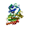
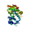
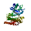
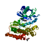
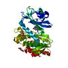
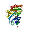
| Title | Crystal structure of haspin in complex with tubercidin | ||||||
 Components Components | Serine/threonine-protein kinase haspin | ||||||
 Keywords Keywords |  TRANSFERASE / kinase / inhibitors / slow off-rate / kinetics / halogen / Structural Genomics / Structural Genomics Consortium / SGC TRANSFERASE / kinase / inhibitors / slow off-rate / kinetics / halogen / Structural Genomics / Structural Genomics Consortium / SGC | ||||||
| Function / homology |  Function and homology information Function and homology informationhistone H3T3 kinase activity / protein localization to chromosome, centromeric region / mitotic sister chromatid cohesion / mitotic spindle assembly checkpoint signaling / spindle / chromosome / mitotic cell cycle / non-specific serine/threonine protein kinase / protein kinase activity / intracellular signal transduction ...histone H3T3 kinase activity / protein localization to chromosome, centromeric region / mitotic sister chromatid cohesion / mitotic spindle assembly checkpoint signaling / spindle / chromosome / mitotic cell cycle / non-specific serine/threonine protein kinase / protein kinase activity / intracellular signal transduction / protein phosphorylation / protein serine kinase activity / centrosome / nucleoplasm / ATP binding / nucleus / cytoplasmSimilarity search - Function | ||||||
| Biological species |  Homo sapiens (human) Homo sapiens (human) | ||||||
| Method | X-RAY DIFFRACTION / SYNCHROTRON / MOLECULAR REPLACEMENT / Resolution: 1.47 Å | ||||||
 Authors Authors | Heroven, C. / Chaikuad, A. / Bountra, C. / Arrowsmith, C.H. / Edwards, A.M. / Knapp, S. / Structural Genomics Consortium (SGC) | ||||||
 Citation Citation | Journal: Angew. Chem. Int. Ed. Engl. / Year: 2018 Title: Halogen-Aromatic pi Interactions Modulate Inhibitor Residence Times. Authors: Heroven, C. / Georgi, V. / Ganotra, G.K. / Brennan, P. / Wolfreys, F. / Wade, R.C. / Fernandez-Montalvan, A.E. / Chaikuad, A. / Knapp, S. | ||||||
| History |
|
- Structure visualization
| Structure viewer | Molecule: MolmilJmol/JSmol |
|---|
- Downloads & links
-Download
| PDBx/mmCIF format | 6g38.cif.gz | 152.5 KB | Display | PDBx/mmCIF format |
|---|---|---|---|---|
| PDB format | pdb6g38.ent.gz | 117.3 KB | Display | PDB format |
| PDBx/mmJSON format | 6g38.json.gz | Tree view | PDBx/mmJSON format | |
| Others |  Other downloads Other downloads |
-Validation report
| Arichive directory | https://data.pdbj.org/pub/pdb/validation_reports/g3/6g38ftp://data.pdbj.org/pub/pdb/validation_reports/g3/6g38 | HTTPS FTP |
|---|
-Related structure data
| Related structure data |  6g33C  6g34C  6g35C  6g36C  6g37C  6g39C  6g3aC  4oucS C: citing same article ( S: Starting model for refinement |
|---|---|
| Similar structure data |


-Links
 PDBj
PDBj- Assembly
| Deposited unit | 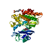
| ||||||||
|---|---|---|---|---|---|---|---|---|---|
| 1 |
| ||||||||
| Unit cell |
|
-Components
-Protein , 1 types, 1 molecules A
| #1: Protein | Mass: 40711.484 Da / Num. of mol.: 1 Source method: isolated from a genetically manipulated source Source: (gene. exp.) Homo sapiens (human) / Gene: HASPIN, GSG2 / Plasmid: pNIC28-Bsa4 / Production host:  Escherichia coli BL21(DE3) (bacteria) / Variant (production host): R3 Escherichia coli BL21(DE3) (bacteria) / Variant (production host): R3References: UniProt: Q8TF76, non-specific serine/threonine protein kinase |
|---|
-Non-polymers , 5 types, 273 molecules 


| #2: Chemical | ChemComp-TBN / ' Mass: 266.253 Da / Num. of mol.: 1 / Source method: obtained synthetically / Formula: C11H14N4O4 Mass: 266.253 Da / Num. of mol.: 1 / Source method: obtained synthetically / Formula: C11H14N4O4 |
|---|---|
| #3: Chemical | ChemComp-DMS / Dimethyl sulfoxide Mass: 78.133 Da / Num. of mol.: 1 / Source method: obtained synthetically / Formula: C2H6OS / Comment: DMSO, precipitant*YM Mass: 78.133 Da / Num. of mol.: 1 / Source method: obtained synthetically / Formula: C2H6OS / Comment: DMSO, precipitant*YM |
| #4: Chemical | ChemComp-EPE / HEPES Mass: 238.305 Da / Num. of mol.: 1 / Source method: obtained synthetically / Formula: C8H18N2O4S / Comment: pH buffer*YM Mass: 238.305 Da / Num. of mol.: 1 / Source method: obtained synthetically / Formula: C8H18N2O4S / Comment: pH buffer*YM |
| #5: Chemical | ChemComp-PO4 / Phosphate Mass: 94.971 Da / Num. of mol.: 1 / Source method: obtained synthetically / Formula: PO4 Mass: 94.971 Da / Num. of mol.: 1 / Source method: obtained synthetically / Formula: PO4 |
| #6: Water | ChemComp-HOH / WaterMass: 18.015 Da / Num. of mol.: 269 / Source method: isolated from a natural source / Formula: H2O |
-Experimental details
-Experiment
| Experiment | Method: X-RAY DIFFRACTION / Number of used crystals: 1 |
|---|
- Sample preparation
| Crystal | Density Matthews: 3.04 Å3/Da / Density % sol: 59.54 % |
|---|---|
| Crystal grow | Temperature: 277.15 K / Method: vapor diffusion, sitting drop / Details: 51-63% MPD and 0.1M SPG buffer, pH 6.0-6.5 / PH range: 6.0-6.5 |
-Data collection
| Diffraction | Mean temperature: 100 K |
|---|---|
| Diffraction source | Source: SYNCHROTRON / Site: Diamond  / Beamline: I02 / Wavelength: 0.9794 Å / Beamline: I02 / Wavelength: 0.9794 Å |
| Detector | Type: DECTRIS PILATUS3 6M / Detector: PIXEL / Date: Nov 30, 2014 |
| Radiation | Protocol: SINGLE WAVELENGTH / Monochromatic (M) / Laue (L): M / Scattering type: x-ray |
| Radiation wavelength | Wavelength: 0.9794 Å / Relative weight: 1 |
| Reflection | Resolution: 1.43→24.05 Å / Num. obs: 92144 / % possible obs: 99.9 % / Redundancy: 5.9 % / CC1/2: 0.994 / Rmerge(I) obs: 0.102 / Net I/σ(I): 8.6 |
| Reflection shell | Resolution: 1.43→1.45 Å / Redundancy: 4.2 % / Mean I/σ(I) obs: 1.2 / Num. unique obs: 4423 / CC1/2: 0.379 / % possible all: 98.3 |
- Processing
| Software |
| ||||||||||||||||||||||||||||||||||||||||||||||||||||||||||||||||||||||||||||||||||||||||||||||||||||||||||||||||||||||||||||||||||||||||||||||||||||||||||||||||||||||||||||||||||||||
|---|---|---|---|---|---|---|---|---|---|---|---|---|---|---|---|---|---|---|---|---|---|---|---|---|---|---|---|---|---|---|---|---|---|---|---|---|---|---|---|---|---|---|---|---|---|---|---|---|---|---|---|---|---|---|---|---|---|---|---|---|---|---|---|---|---|---|---|---|---|---|---|---|---|---|---|---|---|---|---|---|---|---|---|---|---|---|---|---|---|---|---|---|---|---|---|---|---|---|---|---|---|---|---|---|---|---|---|---|---|---|---|---|---|---|---|---|---|---|---|---|---|---|---|---|---|---|---|---|---|---|---|---|---|---|---|---|---|---|---|---|---|---|---|---|---|---|---|---|---|---|---|---|---|---|---|---|---|---|---|---|---|---|---|---|---|---|---|---|---|---|---|---|---|---|---|---|---|---|---|---|---|---|---|
| Refinement | Method to determine structure: MOLECULAR REPLACEMENT Starting model: 4OUC Resolution: 1.47→24.05 Å / Cor.coef. Fo:Fc: 0.977 / Cor.coef. Fo:Fc free: 0.974 / SU B: 1.797 / SU ML: 0.033 / Cross valid method: THROUGHOUT / ESU R: 0.049 / ESU R Free: 0.047 / Details: HYDROGENS HAVE BEEN ADDED IN THE RIDING POSITIONS
| ||||||||||||||||||||||||||||||||||||||||||||||||||||||||||||||||||||||||||||||||||||||||||||||||||||||||||||||||||||||||||||||||||||||||||||||||||||||||||||||||||||||||||||||||||||||
| Solvent computation | Ion probe radii: 0.8 Å / Shrinkage radii: 0.8 Å / VDW probe radii: 1.2 Å | ||||||||||||||||||||||||||||||||||||||||||||||||||||||||||||||||||||||||||||||||||||||||||||||||||||||||||||||||||||||||||||||||||||||||||||||||||||||||||||||||||||||||||||||||||||||
| Displacement parameters | Biso mean: 22.281 Å2
| ||||||||||||||||||||||||||||||||||||||||||||||||||||||||||||||||||||||||||||||||||||||||||||||||||||||||||||||||||||||||||||||||||||||||||||||||||||||||||||||||||||||||||||||||||||||
| Refinement step | Cycle: 1 / Resolution: 1.47→24.05 Å
| ||||||||||||||||||||||||||||||||||||||||||||||||||||||||||||||||||||||||||||||||||||||||||||||||||||||||||||||||||||||||||||||||||||||||||||||||||||||||||||||||||||||||||||||||||||||
| Refine LS restraints |
|