 Movie
Movie Controller

[English] 日本語
 Yorodumi
- PDB-7avq: Crystal structure of haspin in complex with disubstituted imidazo... -
+ Open data

- Basic information
| Entry | Database: PDB / ID: 7avq | ||||||
|---|---|---|---|---|---|---|---|
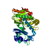
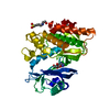
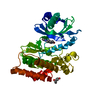
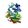
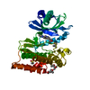
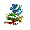
| Title | Crystal structure of haspin in complex with disubstituted imidazo[1,2- b]pyridazine inhibitor (compound 12) | ||||||
 Components Components | Serine/threonine-protein kinase haspin | ||||||
 Keywords Keywords |  TRANSFERASE / kinase / haspin / GSG2 / inhibitor / Structural Genomics / Structural Genomics Consortium / SGC TRANSFERASE / kinase / haspin / GSG2 / inhibitor / Structural Genomics / Structural Genomics Consortium / SGC | ||||||
| Function / homology |  Function and homology information Function and homology informationhistone H3T3 kinase activity / protein localization to chromosome, centromeric region / mitotic sister chromatid cohesion / mitotic spindle assembly checkpoint signaling / spindle / chromosome / mitotic cell cycle / non-specific serine/threonine protein kinase / protein kinase activity / intracellular signal transduction ...histone H3T3 kinase activity / protein localization to chromosome, centromeric region / mitotic sister chromatid cohesion / mitotic spindle assembly checkpoint signaling / spindle / chromosome / mitotic cell cycle / non-specific serine/threonine protein kinase / protein kinase activity / intracellular signal transduction / protein phosphorylation / protein serine kinase activity / centrosome / nucleoplasm / ATP binding / nucleus / cytoplasmSimilarity search - Function | ||||||
| Biological species |  Homo sapiens (human) Homo sapiens (human) | ||||||
| Method | X-RAY DIFFRACTION / SYNCHROTRON / MOLECULAR REPLACEMENT / Resolution: 1.65 Å | ||||||
 Authors Authors | Chaikuad, A. / Bonnet, P. / Routier, S. / Knapp, S. / Structural Genomics Consortium (SGC) | ||||||
 Citation Citation | Journal: J Enzyme Inhib Med Chem / Year: 2020 Title: Design of new disubstituted imidazo[1,2- b ]pyridazine derivatives as selective Haspin inhibitors. Synthesis, binding mode and anticancer biological evaluation. Authors: Elie, J. / Feizbakhsh, O. / Desban, N. / Josselin, B. / Baratte, B. / Bescond, A. / Duez, J. / Fant, X. / Bach, S. / Marie, D. / Place, M. / Ben Salah, S. / Chartier, A. / Berteina-Raboin, S. ...Authors: Elie, J. / Feizbakhsh, O. / Desban, N. / Josselin, B. / Baratte, B. / Bescond, A. / Duez, J. / Fant, X. / Bach, S. / Marie, D. / Place, M. / Ben Salah, S. / Chartier, A. / Berteina-Raboin, S. / Chaikuad, A. / Knapp, S. / Carles, F. / Bonnet, P. / Buron, F. / Routier, S. / Ruchaud, S. | ||||||
| History |
|
- Structure visualization
| Structure viewer | Molecule: MolmilJmol/JSmol |
|---|
- Downloads & links
-Download
| PDBx/mmCIF format | 7avq.cif.gz | 157.7 KB | Display | PDBx/mmCIF format |
|---|---|---|---|---|
| PDB format | pdb7avq.ent.gz | 122.7 KB | Display | PDB format |
| PDBx/mmJSON format | 7avq.json.gz | Tree view | PDBx/mmJSON format | |
| Others |  Other downloads Other downloads |
-Validation report
| Arichive directory | https://data.pdbj.org/pub/pdb/validation_reports/av/7avqftp://data.pdbj.org/pub/pdb/validation_reports/av/7avq | HTTPS FTP |
|---|
-Related structure data
| Related structure data |  4oucS S: Starting model for refinement |
|---|---|
| Similar structure data |


-Links
 PDBj
PDBj- Assembly
| Deposited unit | 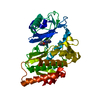
| ||||||||
|---|---|---|---|---|---|---|---|---|---|
| 1 |
| ||||||||
| Unit cell |
|
-Components
-Protein , 1 types, 1 molecules A
| #1: Protein | Mass: 40711.484 Da / Num. of mol.: 1 Source method: isolated from a genetically manipulated source Source: (gene. exp.) Homo sapiens (human) / Gene: HASPIN, GSG2 / Plasmid: pNIC28-Bsa4 / Production host:  Escherichia coli BL21(DE3) (bacteria) / Variant (production host): -R3-pRARE2 Escherichia coli BL21(DE3) (bacteria) / Variant (production host): -R3-pRARE2References: UniProt: Q8TF76, non-specific serine/threonine protein kinase |
|---|
-Non-polymers , 5 types, 299 molecules 


| #2: Chemical | ChemComp-S1Z / ( Mass: 322.364 Da / Num. of mol.: 1 / Source method: obtained synthetically / Formula: C17H18N6O / Feature type: SUBJECT OF INVESTIGATION Mass: 322.364 Da / Num. of mol.: 1 / Source method: obtained synthetically / Formula: C17H18N6O / Feature type: SUBJECT OF INVESTIGATION | ||||
|---|---|---|---|---|---|
| #3: Chemical | ChemComp-NA /  Mass: 22.990 Da / Num. of mol.: 1 / Source method: obtained synthetically / Formula: Na Mass: 22.990 Da / Num. of mol.: 1 / Source method: obtained synthetically / Formula: Na | ||||
| #4: Chemical | Glycerol Mass: 92.094 Da / Num. of mol.: 2 / Source method: obtained synthetically / Formula: C3H8O3 Mass: 92.094 Da / Num. of mol.: 2 / Source method: obtained synthetically / Formula: C3H8O3#5: Chemical | ChemComp-MPD / ( | 2-Methyl-2,4-pentanediol Mass: 118.174 Da / Num. of mol.: 1 / Source method: obtained synthetically / Formula: C6H14O2 / Comment: precipitant*YM Mass: 118.174 Da / Num. of mol.: 1 / Source method: obtained synthetically / Formula: C6H14O2 / Comment: precipitant*YM#6: Water | ChemComp-HOH / | WaterMass: 18.015 Da / Num. of mol.: 294 / Source method: isolated from a natural source / Formula: H2O |
-Details
| Has ligand of interest | Y |
|---|
-Experimental details
-Experiment
| Experiment | Method: X-RAY DIFFRACTION / Number of used crystals: 1 |
|---|
- Sample preparation
| Crystal | Density Matthews: 2.9 Å3/Da / Density % sol: 57.63 % |
|---|---|
| Crystal grow | Temperature: 277.15 K / Method: vapor diffusion, sitting drop / Details: 63% MPD and 0.1 M SPG, pH 6.5 |
-Data collection
| Diffraction | Mean temperature: 100 K / Serial crystal experiment: N | ||||||||||||||||||||||||||||||||||||||||||||||||||||||||||||||||||||||||||||||||||||||||||||||||||||||||||||||
|---|---|---|---|---|---|---|---|---|---|---|---|---|---|---|---|---|---|---|---|---|---|---|---|---|---|---|---|---|---|---|---|---|---|---|---|---|---|---|---|---|---|---|---|---|---|---|---|---|---|---|---|---|---|---|---|---|---|---|---|---|---|---|---|---|---|---|---|---|---|---|---|---|---|---|---|---|---|---|---|---|---|---|---|---|---|---|---|---|---|---|---|---|---|---|---|---|---|---|---|---|---|---|---|---|---|---|---|---|---|---|---|
| Diffraction source | Source: SYNCHROTRON / Site: Diamond  / Beamline: I03 / Wavelength: 0.97626 Å / Beamline: I03 / Wavelength: 0.97626 Å | ||||||||||||||||||||||||||||||||||||||||||||||||||||||||||||||||||||||||||||||||||||||||||||||||||||||||||||||
| Detector | Type: DECTRIS PILATUS3 6M / Detector: PIXEL / Date: Dec 18, 2015 | ||||||||||||||||||||||||||||||||||||||||||||||||||||||||||||||||||||||||||||||||||||||||||||||||||||||||||||||
| Radiation | Protocol: SINGLE WAVELENGTH / Monochromatic (M) / Laue (L): M / Scattering type: x-ray | ||||||||||||||||||||||||||||||||||||||||||||||||||||||||||||||||||||||||||||||||||||||||||||||||||||||||||||||
| Radiation wavelength | Wavelength: 0.97626 Å / Relative weight: 1 | ||||||||||||||||||||||||||||||||||||||||||||||||||||||||||||||||||||||||||||||||||||||||||||||||||||||||||||||
| Reflection | Resolution: 1.65→57.83 Å / Num. obs: 56638 / % possible obs: 98.3 % / Redundancy: 5.3 % / CC1/2: 0.998 / Rmerge(I) obs: 0.067 / Rpim(I) all: 0.031 / Rrim(I) all: 0.074 / Net I/av σ(I): 4.8 / Net I/σ(I): 11.9 | ||||||||||||||||||||||||||||||||||||||||||||||||||||||||||||||||||||||||||||||||||||||||||||||||||||||||||||||
| Reflection shell | Diffraction-ID: 1
|
- Processing
| Software |
| ||||||||||||||||||||||||||||||||||||||||||||||||||||||||||||
|---|---|---|---|---|---|---|---|---|---|---|---|---|---|---|---|---|---|---|---|---|---|---|---|---|---|---|---|---|---|---|---|---|---|---|---|---|---|---|---|---|---|---|---|---|---|---|---|---|---|---|---|---|---|---|---|---|---|---|---|---|---|
| Refinement | Method to determine structure: MOLECULAR REPLACEMENT Starting model: 4OUC Resolution: 1.65→57.83 Å / Cor.coef. Fo:Fc: 0.975 / Cor.coef. Fo:Fc free: 0.969 / SU B: 3.336 / SU ML: 0.056 / SU R Cruickshank DPI: 0.0725 / Cross valid method: THROUGHOUT / σ(F): 0 / ESU R: 0.073 / ESU R Free: 0.072 / Stereochemistry target values: MAXIMUM LIKELIHOOD Details: U VALUES : WITH TLS ADDED HYDROGENS HAVE BEEN ADDED IN THE RIDING POSITIONS
| ||||||||||||||||||||||||||||||||||||||||||||||||||||||||||||
| Solvent computation | Ion probe radii: 0.8 Å / Shrinkage radii: 0.8 Å / VDW probe radii: 1.2 Å / Solvent model: MASK | ||||||||||||||||||||||||||||||||||||||||||||||||||||||||||||
| Displacement parameters | Biso max: 81.47 Å2 / Biso mean: 32.098 Å2 / Biso min: 17.1 Å2
| ||||||||||||||||||||||||||||||||||||||||||||||||||||||||||||
| Refinement step | Cycle: final / Resolution: 1.65→57.83 Å
| ||||||||||||||||||||||||||||||||||||||||||||||||||||||||||||
| Refine LS restraints |
| ||||||||||||||||||||||||||||||||||||||||||||||||||||||||||||
| LS refinement shell | Resolution: 1.65→1.693 Å / Rfactor Rfree error: 0 / Total num. of bins used: 20
| ||||||||||||||||||||||||||||||||||||||||||||||||||||||||||||
| Refinement TLS params. | Method: refined / Origin x: 80.5891 Å / Origin y: 72.0721 Å / Origin z: 35.1926 Å
|
