 Movie
Movie Controller


+ Open data

- Basic information
| Entry | Database: PDB / ID: 6mj8 | ||||||
|---|---|---|---|---|---|---|---|
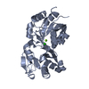
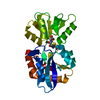
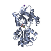
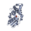
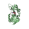
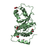
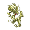
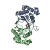
| Title | Structure of Candida glabrata Csm1:Mam1 complex | ||||||
 Components Components |
| ||||||
 Keywords Keywords |  CELL CYCLE / monopolin / kinetochore CELL CYCLE / monopolin / kinetochore | ||||||
| Function / homology |  Function and homology informationmonopolin complex / spindle attachment to meiosis I kinetochore / protein localization to nucleolar rDNA repeats / meiotic sister chromatid cohesion, centromeric / rDNA chromatin condensation / nuclear envelope / nucleolus / identical protein binding Function and homology informationmonopolin complex / spindle attachment to meiosis I kinetochore / protein localization to nucleolar rDNA repeats / meiotic sister chromatid cohesion, centromeric / rDNA chromatin condensation / nuclear envelope / nucleolus / identical protein bindingSimilarity search - Function | ||||||
| Biological species |  Candida glabrata (fungus) Candida glabrata (fungus) | ||||||
| Method | X-RAY DIFFRACTION / SYNCHROTRON / MOLECULAR REPLACEMENT / Resolution: 3.03 Å | ||||||
 Authors Authors | Singh, N. / Corbett, K.D. | ||||||
| Funding support |  United States, 1items United States, 1items
| ||||||
 Citation Citation | Journal: Chromosoma / Year: 2019 Title: The molecular basis of monopolin recruitment to the kinetochore. Authors: Plowman, R. / Singh, N. / Tromer, E.C. / Payan, A. / Duro, E. / Spanos, C. / Rappsilber, J. / Snel, B. / Kops, G.J.P.L. / Corbett, K.D. / Marston, A.L. | ||||||
| History |
|
- Structure visualization
| Structure viewer | Molecule: MolmilJmol/JSmol |
|---|
- Downloads & links
-Download
| PDBx/mmCIF format | 6mj8.cif.gz | 111 KB | Display | PDBx/mmCIF format |
|---|---|---|---|---|
| PDB format | pdb6mj8.ent.gz | 83.9 KB | Display | PDB format |
| PDBx/mmJSON format | 6mj8.json.gz | Tree view | PDBx/mmJSON format | |
| Others |  Other downloads Other downloads |
-Validation report
| Arichive directory | https://data.pdbj.org/pub/pdb/validation_reports/mj/6mj8ftp://data.pdbj.org/pub/pdb/validation_reports/mj/6mj8 | HTTPS FTP |
|---|
-Related structure data
| Related structure data | 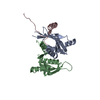 6mjbC 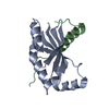 6mjcC 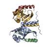 6mjeC 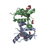 3n4rS S: Starting model for refinement C: citing same article ( |
|---|---|
| Similar structure data | |
| Experimental dataset #1 | Data reference: 10.15785/SBGRID/607 / Data set type: diffraction image data / Details: SBGrid |


-Links
 PDBj
PDBj- Assembly
| Deposited unit | 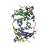
| ||||||||
|---|---|---|---|---|---|---|---|---|---|
| 1 |
| ||||||||
| Unit cell |
|
-Components
| #1: Protein | Mass: 13241.077 Da / Num. of mol.: 2 / Fragment: UNP residues 69-181 Source method: isolated from a genetically manipulated source Source: (gene. exp.) Candida glabrata (fungus) / Gene: AO440_000897, AO440_004693 / Production host:  Escherichia coli (E. coli) / References: UniProt: A0A0W0CH22, UniProt: Q6FVN3*PLUS Escherichia coli (E. coli) / References: UniProt: A0A0W0CH22, UniProt: Q6FVN3*PLUS#2: Protein | Mass: 6611.495 Da / Num. of mol.: 2 / Fragment: UNP residues 162-216 Source method: isolated from a genetically manipulated source Source: (gene. exp.) Candida glabrata (fungus) / Gene: CAGL0G03377g / Production host: Escherichia coli (E. coli) / References: UniProt: Q6FTD4 |
|---|
-Experimental details
-Experiment
| Experiment | Method: X-RAY DIFFRACTION / Number of used crystals: 1 |
|---|
- Sample preparation
| Crystal | Density Matthews: 2.03 Å3/Da / Density % sol: 39.49 % |
|---|---|
| Crystal grow | Temperature: 293 K / Method: vapor diffusion, hanging drop / pH: 6.5 Details: 0.1 M MES, pH 6.5, 0.6 M sodium chloride, 20% PEG4000, cryoprotectant: 20% PEG400 |
-Data collection
| Diffraction | Mean temperature: 100 K |
|---|---|
| Diffraction source | Source: SYNCHROTRON / Site: APS / Beamline: 24-ID-E / Wavelength: 0.97918 Å |
| Detector | Type: ADSC QUANTUM 315 / Detector: CCD / Date: Dec 5, 2013 |
| Radiation | Monochromator: Cryogenically-cooled single crystal Si(220) side bounce Protocol: SINGLE WAVELENGTH / Monochromatic (M) / Laue (L): M / Scattering type: x-ray |
| Radiation wavelength | Wavelength: 0.97918 Å / Relative weight: 1 |
| Reflection | Resolution: 3.03→55 Å / Num. obs: 6723 / % possible obs: 100 % / Redundancy: 6.9 % / Rmerge(I) obs: 0.308 / Rpim(I) all: 0.136 / Rrim(I) all: 0.358 / Net I/σ(I): 8.4 |
| Reflection shell | Resolution: 3.03→3.2 Å / Rmerge(I) obs: 1.651 / Num. unique obs: 958 / Rpim(I) all: 0.714 / Rrim(I) all: 1.919 |
- Processing
| Software |
| ||||||||||||||||||||||||||||||||||||||||
|---|---|---|---|---|---|---|---|---|---|---|---|---|---|---|---|---|---|---|---|---|---|---|---|---|---|---|---|---|---|---|---|---|---|---|---|---|---|---|---|---|---|
| Refinement | Method to determine structure: MOLECULAR REPLACEMENT Starting model: PDB entry 3N4R Resolution: 3.03→54.915 Å / SU ML: 0.1 / Cross valid method: FREE R-VALUE / σ(F): 1.34 / Phase error: 29.6
| ||||||||||||||||||||||||||||||||||||||||
| Solvent computation | Shrinkage radii: 0.9 Å / VDW probe radii: 1.11 Å | ||||||||||||||||||||||||||||||||||||||||
| Refinement step | Cycle: LAST / Resolution: 3.03→54.915 Å
| ||||||||||||||||||||||||||||||||||||||||
| Refine LS restraints |
| ||||||||||||||||||||||||||||||||||||||||
| LS refinement shell |
| ||||||||||||||||||||||||||||||||||||||||
| Refinement TLS params. | Method: refined / Origin x: 6.4084 Å / Origin y: 7.5734 Å / Origin z: 12.409 Å
| ||||||||||||||||||||||||||||||||||||||||
| Refinement TLS group | Selection details: all |
