 Movie
Movie Controller

[English] 日本語
 Yorodumi
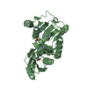
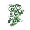
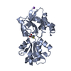
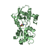
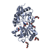
- PDB-4eq9: 1.4 Angstrom Crystal Structure of ABC Transporter Glutathione-Bin... -
+ Open data

- Basic information
| Entry | Database: PDB / ID: 4eq9 | ||||||
|---|---|---|---|---|---|---|---|
| Title | 1.4 Angstrom Crystal Structure of ABC Transporter Glutathione-Binding Protein GshT from Streptococcus pneumoniae strain Canada MDR_19A in Complex with Glutathione | ||||||
 Components Components | ABC transporter substrate-binding protein-amino acid transport | ||||||
 Keywords Keywords |  TRANSPORT PROTEIN / Structural Genomics / NIAID / National Institute of Allergy and Infectious Diseases / Center for Structural Genomics of Infectious Diseases / CSGID TRANSPORT PROTEIN / Structural Genomics / NIAID / National Institute of Allergy and Infectious Diseases / Center for Structural Genomics of Infectious Diseases / CSGID | ||||||
| Function / homology | Bacterial periplasmic substrate-binding proteins / Bacterial extracellular solute-binding proteins, family 3 / Solute-binding protein family 3/N-terminal domain of MltF / Periplasmic binding protein-like II / D-Maltodextrin-Binding Protein; domain 2 / 3-Layer(aba) Sandwich / Alpha Beta / GLUTATHIONE / ABC transporter substrate-binding protein-amino acid transport Function and homology information Function and homology information | ||||||
| Biological species |   Streptococcus pneumoniae (bacteria) Streptococcus pneumoniae (bacteria) | ||||||
| Method | X-RAY DIFFRACTION / SYNCHROTRON / MOLECULAR REPLACEMENT / Resolution: 1.4 Å | ||||||
 Authors Authors | Minasov, G. / Wawrzak, Z. / Stogios, P.J. / Light, S.H. / Kudritska, M. / Yim, V. / Savchenko, A. / Anderson, W.F. / Center for Structural Genomics of Infectious Diseases (CSGID) | ||||||
 Citation Citation | Journal: TO BE PUBLISHED Title: 1.4 Angstrom Crystal Structure of Putative ABC Transporter Substrate-Binding Protein from Streptococcus pneumoniae strain Canada MDR_19A in Complex with Glutathione. Authors: Minasov, G. / Wawrzak, Z. / Stogios, P.J. / Light, S.H. / Kudritska, M. / Yim, V. / Savchenko, A. / Anderson, W.F. / Center for Structural Genomics of Infectious Diseases (CSGID) | ||||||
| History |
|
- Structure visualization
| Structure viewer | Molecule: MolmilJmol/JSmol |
|---|
- Downloads & links
-Download
| PDBx/mmCIF format | 4eq9.cif.gz | 132.9 KB | Display | PDBx/mmCIF format |
|---|---|---|---|---|
| PDB format | pdb4eq9.ent.gz | 105.3 KB | Display | PDB format |
| PDBx/mmJSON format | 4eq9.json.gz | Tree view | PDBx/mmJSON format | |
| Others |  Other downloads Other downloads |
-Validation report
| Arichive directory | https://data.pdbj.org/pub/pdb/validation_reports/eq/4eq9ftp://data.pdbj.org/pub/pdb/validation_reports/eq/4eq9 | HTTPS FTP |
|---|
-Related structure data
| Related structure data |  2q2aS S: Starting model for refinement |
|---|---|
| Similar structure data | |
| Other databases |


-Links
 PDBj
PDBj

- Assembly
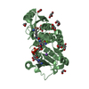
| Deposited unit | 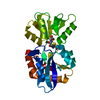
| ||||||||
|---|---|---|---|---|---|---|---|---|---|
| 1 |
| ||||||||
| Unit cell |
|
-Components
| #1: Protein | Mass: 27728.049 Da / Num. of mol.: 1 / Fragment: UNP residues 34-276 Source method: isolated from a genetically manipulated source Source: (gene. exp.) Streptococcus pneumoniae (bacteria) / Strain: Canada MDR_19A / Gene: ABC-SBP, spr0146 / Plasmid: pMCSG7 / Production host: Escherichia coli (E. coli) / Strain (production host): BL21 magic / References: UniProt: Q8DRG2 |
|---|---|
| #2: Chemical | ChemComp-GSH / Glutathione  Mass: 307.323 Da / Num. of mol.: 1 / Source method: obtained synthetically / Formula: C10H17N3O6S Mass: 307.323 Da / Num. of mol.: 1 / Source method: obtained synthetically / Formula: C10H17N3O6S |
| #3: Water | ChemComp-HOH / Water Mass: 18.015 Da / Num. of mol.: 308 / Source method: isolated from a natural source / Formula: H2O Mass: 18.015 Da / Num. of mol.: 308 / Source method: isolated from a natural source / Formula: H2O |
-Experimental details
-Experiment
| Experiment | Method: X-RAY DIFFRACTION / Number of used crystals: 1 |
|---|
- Sample preparation
| Crystal | Density Matthews: 2.36 Å3/Da / Density % sol: 47.98 % |
|---|---|
| Crystal grow | Temperature: 295 K / Method: vapor diffusion, hanging drop / pH: 7.5 Details: Protein: 0.3M Sodium chloride, 0.01M HEPES pH 7.5; Screen: 0.05M ADA (N-(2-Acetamido)iminodiacetic Acid) pH 7.0, 25% (w/v) PEG 3350; Cryo: paratone, VAPOR DIFFUSION, HANGING DROP, temperature 295K |
-Data collection
| Diffraction | Mean temperature: 100 K |
|---|---|
| Diffraction source | Source: SYNCHROTRON / Site: APS  / Beamline: 21-ID-G / Wavelength: 0.97856 Å / Beamline: 21-ID-G / Wavelength: 0.97856 Å |
| Detector | Type: MARMOSAIC 300 mm CCD / Detector: CCD / Date: Apr 12, 2012 / Details: Beryllium lenses |
| Radiation | Monochromator: Diamond / Protocol: SINGLE WAVELENGTH / Monochromatic (M) / Laue (L): M / Scattering type: x-ray |
| Radiation wavelength | Wavelength: 0.97856 Å / Relative weight: 1 |
| Reflection | Resolution: 1.4→30 Å / Num. all: 51214 / Num. obs: 51214 / % possible obs: 99.9 % / Observed criterion σ(I): -3 / Redundancy: 4.3 % / Biso Wilson estimate: 16.3 Å2 / Rmerge(I) obs: 0.043 / Net I/σ(I): 28.9 |
| Reflection shell | Resolution: 1.4→1.42 Å / Redundancy: 4.2 % / Rmerge(I) obs: 0.636 / Mean I/σ(I) obs: 2.2 / Num. unique all: 2565 / % possible all: 100 |
- Processing
| Software |
| |||||||||||||||||||||||||||||||||||||||||||||||||||||||||||||||||||||||||||||||||||||||||||||||||||||||||||||||||||||||||||||||||||||||||||||||||||||||||||||||||||||||||||||||||||||||||||||||||||||||||||||||||||||||||||||||||
|---|---|---|---|---|---|---|---|---|---|---|---|---|---|---|---|---|---|---|---|---|---|---|---|---|---|---|---|---|---|---|---|---|---|---|---|---|---|---|---|---|---|---|---|---|---|---|---|---|---|---|---|---|---|---|---|---|---|---|---|---|---|---|---|---|---|---|---|---|---|---|---|---|---|---|---|---|---|---|---|---|---|---|---|---|---|---|---|---|---|---|---|---|---|---|---|---|---|---|---|---|---|---|---|---|---|---|---|---|---|---|---|---|---|---|---|---|---|---|---|---|---|---|---|---|---|---|---|---|---|---|---|---|---|---|---|---|---|---|---|---|---|---|---|---|---|---|---|---|---|---|---|---|---|---|---|---|---|---|---|---|---|---|---|---|---|---|---|---|---|---|---|---|---|---|---|---|---|---|---|---|---|---|---|---|---|---|---|---|---|---|---|---|---|---|---|---|---|---|---|---|---|---|---|---|---|---|---|---|---|---|---|---|---|---|---|---|---|---|---|---|---|---|---|---|---|---|
| Refinement | Method to determine structure: MOLECULAR REPLACEMENT Starting model: 2Q2A Resolution: 1.4→27.03 Å / Cor.coef. Fo:Fc: 0.969 / Cor.coef. Fo:Fc free: 0.958 / SU B: 2.17 / SU ML: 0.039 Isotropic thermal model: Thermal Factors Individually Refined Cross valid method: THROUGHOUT / ESU R: 0.06 / ESU R Free: 0.061 / Stereochemistry target values: MAXIMUM LIKELIHOOD / Details: HYDROGENS HAVE BEEN ADDED IN THE RIDING POSITIONS
| |||||||||||||||||||||||||||||||||||||||||||||||||||||||||||||||||||||||||||||||||||||||||||||||||||||||||||||||||||||||||||||||||||||||||||||||||||||||||||||||||||||||||||||||||||||||||||||||||||||||||||||||||||||||||||||||||
| Solvent computation | Ion probe radii: 0.8 Å / Shrinkage radii: 0.8 Å / VDW probe radii: 1.2 Å / Solvent model: MASK | |||||||||||||||||||||||||||||||||||||||||||||||||||||||||||||||||||||||||||||||||||||||||||||||||||||||||||||||||||||||||||||||||||||||||||||||||||||||||||||||||||||||||||||||||||||||||||||||||||||||||||||||||||||||||||||||||
| Displacement parameters | Biso mean: 16.692 Å2
| |||||||||||||||||||||||||||||||||||||||||||||||||||||||||||||||||||||||||||||||||||||||||||||||||||||||||||||||||||||||||||||||||||||||||||||||||||||||||||||||||||||||||||||||||||||||||||||||||||||||||||||||||||||||||||||||||
| Refinement step | Cycle: LAST / Resolution: 1.4→27.03 Å
| |||||||||||||||||||||||||||||||||||||||||||||||||||||||||||||||||||||||||||||||||||||||||||||||||||||||||||||||||||||||||||||||||||||||||||||||||||||||||||||||||||||||||||||||||||||||||||||||||||||||||||||||||||||||||||||||||
| Refine LS restraints |
| |||||||||||||||||||||||||||||||||||||||||||||||||||||||||||||||||||||||||||||||||||||||||||||||||||||||||||||||||||||||||||||||||||||||||||||||||||||||||||||||||||||||||||||||||||||||||||||||||||||||||||||||||||||||||||||||||
| LS refinement shell | Resolution: 1.4→1.436 Å / Total num. of bins used: 20
| |||||||||||||||||||||||||||||||||||||||||||||||||||||||||||||||||||||||||||||||||||||||||||||||||||||||||||||||||||||||||||||||||||||||||||||||||||||||||||||||||||||||||||||||||||||||||||||||||||||||||||||||||||||||||||||||||
| Refinement TLS params. | Method: refined / Refine-ID: X-RAY DIFFRACTION
| |||||||||||||||||||||||||||||||||||||||||||||||||||||||||||||||||||||||||||||||||||||||||||||||||||||||||||||||||||||||||||||||||||||||||||||||||||||||||||||||||||||||||||||||||||||||||||||||||||||||||||||||||||||||||||||||||
| Refinement TLS group |
|
