 Movie
Movie Controller


+ Open data

- Basic information
| Entry | Database: PDB / ID: 6j53 | |||||||||
|---|---|---|---|---|---|---|---|---|---|---|
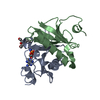
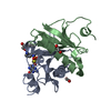
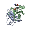
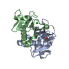
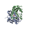
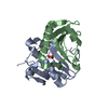
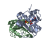
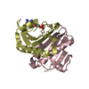
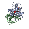
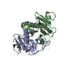
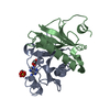
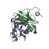
| Title | Crystal structure of human HINT1 complexing with ATP | |||||||||
 Components Components | Histidine triad nucleotide-binding protein 1 | |||||||||
 Keywords Keywords |  HYDROLASE / NUCLEOTIDE BINDING / REGULATION OF TRANSCRIPTION / SIGNAL TRANSDUCTION HYDROLASE / NUCLEOTIDE BINDING / REGULATION OF TRANSCRIPTION / SIGNAL TRANSDUCTION | |||||||||
| Function / homology |  Function and homology information Function and homology informationpurine ribonucleotide catabolic process / Hydrolases; Acting on phosphorus-nitrogen bonds / adenosine 5'-monophosphoramidase activity / deSUMOylase activity / protein desumoylation / histone deacetylase complex / intrinsic apoptotic signaling pathway by p53 class mediator / positive regulation of calcium-mediated signaling / protein kinase C binding / Hydrolases; Acting on peptide bonds (peptidases); Cysteine endopeptidases ...purine ribonucleotide catabolic process / Hydrolases; Acting on phosphorus-nitrogen bonds / adenosine 5'-monophosphoramidase activity / deSUMOylase activity / protein desumoylation / histone deacetylase complex / intrinsic apoptotic signaling pathway by p53 class mediator / positive regulation of calcium-mediated signaling / protein kinase C binding / Hydrolases; Acting on peptide bonds (peptidases); Cysteine endopeptidases / cytoskeleton / hydrolase activity / nucleotide binding / regulation of DNA-templated transcription / signal transduction / proteolysis / extracellular exosome / nucleoplasm / nucleus / plasma membrane / cytosol / cytoplasmSimilarity search - Function | |||||||||
| Biological species |  Homo sapiens (human) Homo sapiens (human) | |||||||||
| Method | X-RAY DIFFRACTION / SYNCHROTRON / MOLECULAR REPLACEMENT / Resolution: 1.52 Å | |||||||||
 Authors Authors | Wang, J. / Fang, P. / Guo, M. | |||||||||
| Funding support |  China, 2items China, 2items
| |||||||||
 Citation Citation | Journal: Nat Commun / Year: 2019 Title: Second messenger Ap4A polymerizes target protein HINT1 to transduce signals in Fc epsilon RI-activated mast cells. Authors: Yu, J. / Liu, Z. / Liang, Y. / Luo, F. / Zhang, J. / Tian, C. / Motzik, A. / Zheng, M. / Kang, J. / Zhong, G. / Liu, C. / Fang, P. / Guo, M. / Razin, E. / Wang, J. | |||||||||
| History |
|
- Structure visualization
| Structure viewer | Molecule: MolmilJmol/JSmol |
|---|
- Downloads & links
-Download
| PDBx/mmCIF format | 6j53.cif.gz | 118.2 KB | Display | PDBx/mmCIF format |
|---|---|---|---|---|
| PDB format | pdb6j53.ent.gz | 88.8 KB | Display | PDB format |
| PDBx/mmJSON format | 6j53.json.gz | Tree view | PDBx/mmJSON format | |
| Others |  Other downloads Other downloads |
-Validation report
| Arichive directory | https://data.pdbj.org/pub/pdb/validation_reports/j5/6j53ftp://data.pdbj.org/pub/pdb/validation_reports/j5/6j53 | HTTPS FTP |
|---|
-Related structure data
| Related structure data |  5ed3C  5ed6C  6j58C 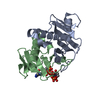 6j5sC  6j5zC 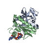 6j64C 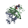 6j65C 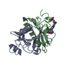 4eqeS S: Starting model for refinement C: citing same article ( |
|---|---|
| Similar structure data |
-Links
 PDBj
PDBj


- Assembly
| Deposited unit | 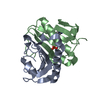
| ||||||||
|---|---|---|---|---|---|---|---|---|---|
| 1 |
| ||||||||
| Unit cell |
|
-Components
| #1: Protein | Mass: 14024.165 Da / Num. of mol.: 2 Source method: isolated from a genetically manipulated source Source: (gene. exp.) Homo sapiens (human) / Gene: HINT1, HINT, PKCI1, PRKCNH1 / Production host:  Escherichia coli (E. coli) / References: UniProt: P49773, Hydrolases Escherichia coli (E. coli) / References: UniProt: P49773, Hydrolases#2: Chemical | ChemComp-AMP / | Adenosine monophosphate  Mass: 347.221 Da / Num. of mol.: 1 / Source method: obtained synthetically / Formula: C10H14N5O7P / Comment: AMP*YM Mass: 347.221 Da / Num. of mol.: 1 / Source method: obtained synthetically / Formula: C10H14N5O7P / Comment: AMP*YM#3: Water | ChemComp-HOH / | Water Mass: 18.015 Da / Num. of mol.: 361 / Source method: isolated from a natural source / Formula: H2O Mass: 18.015 Da / Num. of mol.: 361 / Source method: isolated from a natural source / Formula: H2O |
|---|
-Experimental details
-Experiment
| Experiment | Method: X-RAY DIFFRACTION / Number of used crystals: 1 |
|---|
- Sample preparation
| Crystal | Density Matthews: 2.08 Å3/Da / Density % sol: 40.74 % |
|---|---|
| Crystal grow | Temperature: 291 K / Method: vapor diffusion, sitting drop / pH: 7.5 / Details: HEPES pH 7.5, PEG 3350 |
-Data collection
| Diffraction | Mean temperature: 100 K / Serial crystal experiment: N |
|---|---|
| Diffraction source | Source: SYNCHROTRON / Site: APS  / Beamline: 21-ID-G / Wavelength: 0.97857 Å / Beamline: 21-ID-G / Wavelength: 0.97857 Å |
| Detector | Type: MARMOSAIC 300 mm CCD / Detector: CCD / Date: Jul 6, 2011 |
| Radiation | Protocol: SINGLE WAVELENGTH / Monochromatic (M) / Laue (L): M / Scattering type: x-ray |
| Radiation wavelength | Wavelength: 0.97857 Å / Relative weight: 1 |
| Reflection | Resolution: 1.52→25 Å / Num. obs: 35501 / % possible obs: 99.9 % / Redundancy: 3.5 % / Biso Wilson estimate: 9.54 Å2 / Rmerge(I) obs: 0.036 / Net I/σ(I): 40.5 |
| Reflection shell | Resolution: 1.52→1.55 Å / Rmerge(I) obs: 0.108 / Num. unique obs: 1255 / % possible all: 99.9 |
- Processing
| Software |
| |||||||||||||||||||||||||||||||||||||||||||||||||||||||||||||||||||||||||||||
|---|---|---|---|---|---|---|---|---|---|---|---|---|---|---|---|---|---|---|---|---|---|---|---|---|---|---|---|---|---|---|---|---|---|---|---|---|---|---|---|---|---|---|---|---|---|---|---|---|---|---|---|---|---|---|---|---|---|---|---|---|---|---|---|---|---|---|---|---|---|---|---|---|---|---|---|---|---|---|
| Refinement | Method to determine structure: MOLECULAR REPLACEMENT Starting model: 4EQE Resolution: 1.52→23.843 Å / SU ML: 0.11 / Cross valid method: THROUGHOUT / σ(F): 1.35 / Phase error: 16.34 / Stereochemistry target values: ML
| |||||||||||||||||||||||||||||||||||||||||||||||||||||||||||||||||||||||||||||
| Solvent computation | Shrinkage radii: 0.9 Å / VDW probe radii: 1.11 Å / Solvent model: FLAT BULK SOLVENT MODEL | |||||||||||||||||||||||||||||||||||||||||||||||||||||||||||||||||||||||||||||
| Displacement parameters | Biso max: 52.73 Å2 / Biso mean: 14.4777 Å2 / Biso min: 4.44 Å2 | |||||||||||||||||||||||||||||||||||||||||||||||||||||||||||||||||||||||||||||
| Refinement step | Cycle: final / Resolution: 1.52→23.843 Å
| |||||||||||||||||||||||||||||||||||||||||||||||||||||||||||||||||||||||||||||
| LS refinement shell | Refine-ID: X-RAY DIFFRACTION / Rfactor Rfree error: 0 / Total num. of bins used: 10
| |||||||||||||||||||||||||||||||||||||||||||||||||||||||||||||||||||||||||||||
| Refinement TLS params. | Method: refined / Origin x: 13.2023 Å / Origin y: 1.0436 Å / Origin z: 16.2709 Å
| |||||||||||||||||||||||||||||||||||||||||||||||||||||||||||||||||||||||||||||
| Refinement TLS group | Selection details: ALL |
