 Movie
Movie Controller

[English] 日本語
 Yorodumi
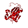
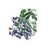
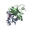
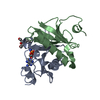
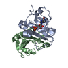
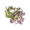
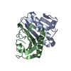
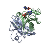
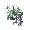
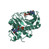
- PDB-4eqe: Crystal structure of histidine triad nucleotide-binding protein 1... -
+ Open data

- Basic information
| Entry | Database: PDB / ID: 4eqe | ||||||
|---|---|---|---|---|---|---|---|
| Title | Crystal structure of histidine triad nucleotide-binding protein 1 (HINT1) from human complexed with Lys-AMS | ||||||
 Components Components | Histidine triad nucleotide-binding protein 1 | ||||||
 Keywords Keywords |  HYDROLASE / HIT domain HYDROLASE / HIT domain | ||||||
| Function / homology |  Function and homology information Function and homology informationpurine ribonucleotide catabolic process / adenylylsulfatase activity / Hydrolases; Acting on phosphorus-nitrogen bonds / adenosine 5'-monophosphoramidase activity / deSUMOylase activity / protein desumoylation / sulfur compound metabolic process / histone deacetylase complex / intrinsic apoptotic signaling pathway by p53 class mediator / positive regulation of calcium-mediated signaling ...purine ribonucleotide catabolic process / adenylylsulfatase activity / Hydrolases; Acting on phosphorus-nitrogen bonds / adenosine 5'-monophosphoramidase activity / deSUMOylase activity / protein desumoylation / sulfur compound metabolic process / histone deacetylase complex / intrinsic apoptotic signaling pathway by p53 class mediator / positive regulation of calcium-mediated signaling / protein kinase C binding / Hydrolases; Acting on peptide bonds (peptidases); Cysteine endopeptidases / cytoskeleton / hydrolase activity / nucleotide binding / regulation of DNA-templated transcription / signal transduction / proteolysis / extracellular exosome / nucleoplasm / nucleus / plasma membrane / cytosol / cytoplasmSimilarity search - Function | ||||||
| Biological species |  Homo sapiens (human) Homo sapiens (human) | ||||||
| Method | X-RAY DIFFRACTION / SYNCHROTRON / MOLECULAR REPLACEMENT / Resolution: 1.52 Å | ||||||
 Authors Authors | Wang, J. / Fang, P. / Guo, M. | ||||||
 Citation Citation | Journal: J.Phys.Chem.B / Year: 2012 Title: Side chain independent recognition of aminoacyl adenylates by the hint1 transcription suppressor. Authors: Wang, J. / Fang, P. / Schimmel, P. / Guo, M. | ||||||
| History |
|
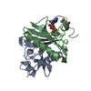
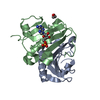
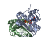
- Structure visualization
| Structure viewer | Molecule: MolmilJmol/JSmol |
|---|
- Downloads & links
-Download
| PDBx/mmCIF format | 4eqe.cif.gz | 117.7 KB | Display | PDBx/mmCIF format |
|---|---|---|---|---|
| PDB format | pdb4eqe.ent.gz | 89.1 KB | Display | PDB format |
| PDBx/mmJSON format | 4eqe.json.gz | Tree view | PDBx/mmJSON format | |
| Others |  Other downloads Other downloads |
-Validation report
| Arichive directory | https://data.pdbj.org/pub/pdb/validation_reports/eq/4eqeftp://data.pdbj.org/pub/pdb/validation_reports/eq/4eqe | HTTPS FTP |
|---|
-Related structure data
| Related structure data |  4eqgC 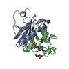 4eqhC  6rhnS C: citing same article ( S: Starting model for refinement |
|---|---|
| Similar structure data |


-Links
 PDBj
PDBj


- Assembly
| Deposited unit | 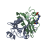
| ||||||||
|---|---|---|---|---|---|---|---|---|---|
| 1 |
| ||||||||
| Unit cell |
| ||||||||
| Components on special symmetry positions |
|
-Components
| #1: Protein | Mass: 14024.165 Da / Num. of mol.: 2 Source method: isolated from a genetically manipulated source Source: (gene. exp.) Homo sapiens (human) / Gene: HINT1, HINT / Production host:  Escherichia coli (E. coli) / References: UniProt: P49773, Hydrolases Escherichia coli (E. coli) / References: UniProt: P49773, Hydrolases#2: Chemical | ChemComp-EPE / | HEPES  Mass: 238.305 Da / Num. of mol.: 1 / Source method: obtained synthetically / Formula: C8H18N2O4S / Comment: pH buffer*YM Mass: 238.305 Da / Num. of mol.: 1 / Source method: obtained synthetically / Formula: C8H18N2O4S / Comment: pH buffer*YM#3: Chemical | ChemComp-KAA / | 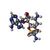  Mass: 474.492 Da / Num. of mol.: 1 / Source method: obtained synthetically / Formula: C16H26N8O7S Mass: 474.492 Da / Num. of mol.: 1 / Source method: obtained synthetically / Formula: C16H26N8O7S#4: Water | ChemComp-HOH / | Water Mass: 18.015 Da / Num. of mol.: 291 / Source method: isolated from a natural source / Formula: H2O Mass: 18.015 Da / Num. of mol.: 291 / Source method: isolated from a natural source / Formula: H2O |
|---|
-Experimental details
-Experiment
| Experiment | Method: X-RAY DIFFRACTION / Number of used crystals: 1 |
|---|
- Sample preparation
| Crystal | Density Matthews: 2.09 Å3/Da / Density % sol: 41.13 % |
|---|---|
| Crystal grow | Temperature: 291 K / Method: vapor diffusion, sitting drop / pH: 7.5 Details: 25-28% PEG3350, 0.1 M HEPES, pH 7.5, VAPOR DIFFUSION, SITTING DROP, temperature 291K |
-Data collection
| Diffraction | Mean temperature: 100 K | |||||||||||||||||||||||||||||||||||||||||||||||||||||||||||||||||||||||||||||
|---|---|---|---|---|---|---|---|---|---|---|---|---|---|---|---|---|---|---|---|---|---|---|---|---|---|---|---|---|---|---|---|---|---|---|---|---|---|---|---|---|---|---|---|---|---|---|---|---|---|---|---|---|---|---|---|---|---|---|---|---|---|---|---|---|---|---|---|---|---|---|---|---|---|---|---|---|---|---|
| Diffraction source | Source: SYNCHROTRON / Site: APS  / Beamline: 21-ID-D / Wavelength: 1.033 Å / Beamline: 21-ID-D / Wavelength: 1.033 Å | |||||||||||||||||||||||||||||||||||||||||||||||||||||||||||||||||||||||||||||
| Detector | Type: MARMOSAIC 300 mm CCD / Detector: CCD / Date: Jul 6, 2011 | |||||||||||||||||||||||||||||||||||||||||||||||||||||||||||||||||||||||||||||
| Radiation | Monochromator: Kohzu Si(111) / Protocol: SINGLE WAVELENGTH / Monochromatic (M) / Laue (L): M / Scattering type: x-ray | |||||||||||||||||||||||||||||||||||||||||||||||||||||||||||||||||||||||||||||
| Radiation wavelength | Wavelength: 1.033 Å / Relative weight: 1 | |||||||||||||||||||||||||||||||||||||||||||||||||||||||||||||||||||||||||||||
| Reflection | Resolution: 1.52→25 Å / Num. obs: 35542 / % possible obs: 99.3 % / Redundancy: 7.5 % / Rmerge(I) obs: 0.048 / Χ2: 0.952 / Net I/σ(I): 13.2 | |||||||||||||||||||||||||||||||||||||||||||||||||||||||||||||||||||||||||||||
| Reflection shell |
|
- Processing
| Software |
| |||||||||||||||||||||||||||||||||||||||||||||||||||||||||||||||||||||||||||||||||||||||||||
|---|---|---|---|---|---|---|---|---|---|---|---|---|---|---|---|---|---|---|---|---|---|---|---|---|---|---|---|---|---|---|---|---|---|---|---|---|---|---|---|---|---|---|---|---|---|---|---|---|---|---|---|---|---|---|---|---|---|---|---|---|---|---|---|---|---|---|---|---|---|---|---|---|---|---|---|---|---|---|---|---|---|---|---|---|---|---|---|---|---|---|---|---|
| Refinement | Method to determine structure: MOLECULAR REPLACEMENT Starting model: PDB ENTRY 6RHN Resolution: 1.52→24.396 Å / Occupancy max: 1 / Occupancy min: 0.28 / FOM work R set: 0.9199 / SU ML: 0.34 / σ(F): 1.34 / Phase error: 14.5 / Stereochemistry target values: ML
| |||||||||||||||||||||||||||||||||||||||||||||||||||||||||||||||||||||||||||||||||||||||||||
| Solvent computation | Shrinkage radii: 0.9 Å / VDW probe radii: 1.11 Å / Solvent model: FLAT BULK SOLVENT MODEL / Bsol: 54.794 Å2 / ksol: 0.381 e/Å3 | |||||||||||||||||||||||||||||||||||||||||||||||||||||||||||||||||||||||||||||||||||||||||||
| Displacement parameters | Biso max: 59.82 Å2 / Biso mean: 16.5139 Å2 / Biso min: 5.68 Å2
| |||||||||||||||||||||||||||||||||||||||||||||||||||||||||||||||||||||||||||||||||||||||||||
| Refinement step | Cycle: LAST / Resolution: 1.52→24.396 Å
| |||||||||||||||||||||||||||||||||||||||||||||||||||||||||||||||||||||||||||||||||||||||||||
| Refine LS restraints |
| |||||||||||||||||||||||||||||||||||||||||||||||||||||||||||||||||||||||||||||||||||||||||||
| LS refinement shell | Refine-ID: X-RAY DIFFRACTION / Total num. of bins used: 12
| |||||||||||||||||||||||||||||||||||||||||||||||||||||||||||||||||||||||||||||||||||||||||||
| Refinement TLS params. | Method: refined / Origin x: 23.5667 Å / Origin y: -0.0215 Å / Origin z: 15.924 Å
| |||||||||||||||||||||||||||||||||||||||||||||||||||||||||||||||||||||||||||||||||||||||||||
| Refinement TLS group |
|
