 Movie
Movie Controller

[English] 日本語
 Yorodumi
- PDB-4zkl: Crystal structure of human histidine triad nucleotide-binding pro... -
+ Open data

- Basic information
| Entry | Database: PDB / ID: 4zkl | ||||||
|---|---|---|---|---|---|---|---|
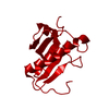
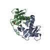
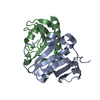
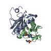
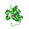
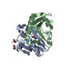
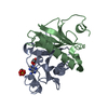
| Title | Crystal structure of human histidine triad nucleotide-binding protein 1 (hHINT1) complexed with JB419 (AP4A analog) | ||||||
 Components Components | Histidine triad nucleotide-binding protein 1 | ||||||
 Keywords Keywords |  HYDROLASE / HINT / HIT / Phosphoramidase / Complex / AP4A analog HYDROLASE / HINT / HIT / Phosphoramidase / Complex / AP4A analog | ||||||
| Function / homology |  Function and homology information Function and homology informationpurine ribonucleotide catabolic process / adenylylsulfatase activity / Hydrolases; Acting on phosphorus-nitrogen bonds / adenosine 5'-monophosphoramidase activity / deSUMOylase activity / protein desumoylation / sulfur compound metabolic process / histone deacetylase complex / intrinsic apoptotic signaling pathway by p53 class mediator / positive regulation of calcium-mediated signaling ...purine ribonucleotide catabolic process / adenylylsulfatase activity / Hydrolases; Acting on phosphorus-nitrogen bonds / adenosine 5'-monophosphoramidase activity / deSUMOylase activity / protein desumoylation / sulfur compound metabolic process / histone deacetylase complex / intrinsic apoptotic signaling pathway by p53 class mediator / positive regulation of calcium-mediated signaling / protein kinase C binding / Hydrolases; Acting on peptide bonds (peptidases); Cysteine endopeptidases / cytoskeleton / hydrolase activity / nucleotide binding / regulation of DNA-templated transcription / signal transduction / proteolysis / extracellular exosome / nucleoplasm / nucleus / plasma membrane / cytosol / cytoplasmSimilarity search - Function | ||||||
| Biological species |  Homo sapiens (human) Homo sapiens (human) | ||||||
| Method | X-RAY DIFFRACTION / SYNCHROTRON / MOLECULAR REPLACEMENT / Resolution: 2.34 Å | ||||||
 Authors Authors | Dolot, R.M. / Kaczmarek, R. / Seda, A. / Krakowiak, A. / Baraniak, J. / Nawrot, B. | ||||||
 Citation Citation | Journal: Int.J.Biol.Macromol. / Year: 2016 Title: Crystallographic studies of the complex of human HINT1 protein with a non-hydrolyzable analog of Ap4A. Authors: Dolot, R. / Kaczmarek, R. / Seda, A. / Krakowiak, A. / Baraniak, J. / Nawrot, B. | ||||||
| History |
|
- Structure visualization
| Structure viewer | Molecule: MolmilJmol/JSmol |
|---|
- Downloads & links
-Download
| PDBx/mmCIF format | 4zkl.cif.gz | 194.1 KB | Display | PDBx/mmCIF format |
|---|---|---|---|---|
| PDB format | pdb4zkl.ent.gz | 156.7 KB | Display | PDB format |
| PDBx/mmJSON format | 4zkl.json.gz | Tree view | PDBx/mmJSON format | |
| Others |  Other downloads Other downloads |
-Validation report
| Arichive directory | https://data.pdbj.org/pub/pdb/validation_reports/zk/4zklftp://data.pdbj.org/pub/pdb/validation_reports/zk/4zkl | HTTPS FTP |
|---|
-Related structure data
| Related structure data |  4zkvC 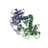 3tw2S S: Starting model for refinement C: citing same article ( |
|---|---|
| Similar structure data |


-Links
 PDBj
PDBj


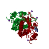
- Assembly
| Deposited unit | 
| ||||||||||||||||||||||||||||||||||||||||||||||||||||||||||||||||||||||||||||||||||||||||||||||||||||||||||||||||||||||||||||||||||||||||||||||||||||||
|---|---|---|---|---|---|---|---|---|---|---|---|---|---|---|---|---|---|---|---|---|---|---|---|---|---|---|---|---|---|---|---|---|---|---|---|---|---|---|---|---|---|---|---|---|---|---|---|---|---|---|---|---|---|---|---|---|---|---|---|---|---|---|---|---|---|---|---|---|---|---|---|---|---|---|---|---|---|---|---|---|---|---|---|---|---|---|---|---|---|---|---|---|---|---|---|---|---|---|---|---|---|---|---|---|---|---|---|---|---|---|---|---|---|---|---|---|---|---|---|---|---|---|---|---|---|---|---|---|---|---|---|---|---|---|---|---|---|---|---|---|---|---|---|---|---|---|---|---|---|---|---|
| 1 | 
| ||||||||||||||||||||||||||||||||||||||||||||||||||||||||||||||||||||||||||||||||||||||||||||||||||||||||||||||||||||||||||||||||||||||||||||||||||||||
| 2 | 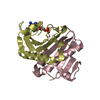
| ||||||||||||||||||||||||||||||||||||||||||||||||||||||||||||||||||||||||||||||||||||||||||||||||||||||||||||||||||||||||||||||||||||||||||||||||||||||
| Unit cell |
| ||||||||||||||||||||||||||||||||||||||||||||||||||||||||||||||||||||||||||||||||||||||||||||||||||||||||||||||||||||||||||||||||||||||||||||||||||||||
| Noncrystallographic symmetry (NCS) | NCS domain:
NCS domain segments: Component-ID: 0 / Beg auth comp-ID: GLY / Beg label comp-ID: GLY / Refine code: 0
NCS ensembles :
|
-Components
| #1: Protein | Mass: 13823.931 Da / Num. of mol.: 4 / Fragment: UNP residues 1-126 Source method: isolated from a genetically manipulated source Source: (gene. exp.) Homo sapiens (human) / Gene: HINT1, HINT, PKCI1, PRKCNH1 / Plasmid: pSGA02 / Production host:  Escherichia coli BL21(DE3) (bacteria) / References: UniProt: P49773, Hydrolases Escherichia coli BL21(DE3) (bacteria) / References: UniProt: P49773, Hydrolases#2: Chemical | Adenosine monophosphate  Mass: 347.221 Da / Num. of mol.: 2 / Source method: obtained synthetically / Formula: C10H14N5O7P / Comment: AMP*YM Mass: 347.221 Da / Num. of mol.: 2 / Source method: obtained synthetically / Formula: C10H14N5O7P / Comment: AMP*YM#3: Chemical | ChemComp-JB6 / ( |   Mass: 796.663 Da / Num. of mol.: 1 / Source method: obtained synthetically / Formula: C24H34N10O13P2S2 Mass: 796.663 Da / Num. of mol.: 1 / Source method: obtained synthetically / Formula: C24H34N10O13P2S2#4: Water | ChemComp-HOH / | Water Mass: 18.015 Da / Num. of mol.: 194 / Source method: isolated from a natural source / Formula: H2O Mass: 18.015 Da / Num. of mol.: 194 / Source method: isolated from a natural source / Formula: H2O |
|---|
-Experimental details
-Experiment
| Experiment | Method: X-RAY DIFFRACTION / Number of used crystals: 1 |
|---|
- Sample preparation
| Crystal | Density Matthews: 3.03 Å3/Da / Density % sol: 59.45 % / Description: plates |
|---|---|
| Crystal grow | Temperature: 281 K / Method: vapor diffusion, hanging drop / pH: 5.5 / Details: 19% w/v PEG4000, 0.1 M sodium cacodylate pH 5.5 |
-Data collection
| Diffraction | Mean temperature: 100 K |
|---|---|
| Diffraction source | Source: SYNCHROTRON / Site: PETRA III, EMBL c/o DESY  / Beamline: P13 (MX1) / Wavelength: 0.9669 Å / Beamline: P13 (MX1) / Wavelength: 0.9669 Å |
| Detector | Type: DECTRIS PILATUS 6M / Detector: PIXEL / Date: Dec 14, 2013 |
| Radiation | Monochromator: Si(111) crystal / Protocol: SINGLE WAVELENGTH / Monochromatic (M) / Laue (L): M / Scattering type: x-ray |
| Radiation wavelength | Wavelength: 0.9669 Å / Relative weight: 1 |
| Reflection | Resolution: 2.34→102.55 Å / Num. obs: 25059 / % possible obs: 97.6 % / Redundancy: 3.3 % / Biso Wilson estimate: 36.7 Å2 / Rmerge(I) obs: 0.096 / Net I/σ(I): 7.1 |
| Reflection shell | Resolution: 2.34→2.42 Å / Redundancy: 3.4 % / Rmerge(I) obs: 0.692 / Mean I/σ(I) obs: 1.6 / % possible all: 99 |
- Processing
| Software |
| ||||||||||||||||||||||||||||||||||||||||||||||||||||||||||||||||||||||||||||||||||||||||||||||||||||||||||||||||||||||||||||||||||||||||||||||||||||||||||||||||||||||||||||||||||||||
|---|---|---|---|---|---|---|---|---|---|---|---|---|---|---|---|---|---|---|---|---|---|---|---|---|---|---|---|---|---|---|---|---|---|---|---|---|---|---|---|---|---|---|---|---|---|---|---|---|---|---|---|---|---|---|---|---|---|---|---|---|---|---|---|---|---|---|---|---|---|---|---|---|---|---|---|---|---|---|---|---|---|---|---|---|---|---|---|---|---|---|---|---|---|---|---|---|---|---|---|---|---|---|---|---|---|---|---|---|---|---|---|---|---|---|---|---|---|---|---|---|---|---|---|---|---|---|---|---|---|---|---|---|---|---|---|---|---|---|---|---|---|---|---|---|---|---|---|---|---|---|---|---|---|---|---|---|---|---|---|---|---|---|---|---|---|---|---|---|---|---|---|---|---|---|---|---|---|---|---|---|---|---|---|
| Refinement | Method to determine structure: MOLECULAR REPLACEMENT Starting model: 3TW2 Resolution: 2.34→102.55 Å / Cor.coef. Fo:Fc: 0.966 / Cor.coef. Fo:Fc free: 0.944 / SU B: 21.98 / SU ML: 0.237 / Cross valid method: THROUGHOUT / ESU R: 0.297 / ESU R Free: 0.229 / Stereochemistry target values: MAXIMUM LIKELIHOOD / Details: HYDROGENS HAVE BEEN ADDED IN THE RIDING POSITIONS
| ||||||||||||||||||||||||||||||||||||||||||||||||||||||||||||||||||||||||||||||||||||||||||||||||||||||||||||||||||||||||||||||||||||||||||||||||||||||||||||||||||||||||||||||||||||||
| Solvent computation | Ion probe radii: 0.8 Å / Shrinkage radii: 0.8 Å / VDW probe radii: 1.2 Å / Solvent model: MASK | ||||||||||||||||||||||||||||||||||||||||||||||||||||||||||||||||||||||||||||||||||||||||||||||||||||||||||||||||||||||||||||||||||||||||||||||||||||||||||||||||||||||||||||||||||||||
| Displacement parameters | Biso mean: 60.124 Å2
| ||||||||||||||||||||||||||||||||||||||||||||||||||||||||||||||||||||||||||||||||||||||||||||||||||||||||||||||||||||||||||||||||||||||||||||||||||||||||||||||||||||||||||||||||||||||
| Refinement step | Cycle: LAST / Resolution: 2.34→102.55 Å
| ||||||||||||||||||||||||||||||||||||||||||||||||||||||||||||||||||||||||||||||||||||||||||||||||||||||||||||||||||||||||||||||||||||||||||||||||||||||||||||||||||||||||||||||||||||||
| Refine LS restraints |
|