 Movie
Movie Controller

[English] 日本語
 Yorodumi
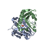
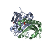
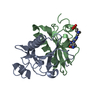
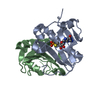
- PDB-5km0: Human Histidine Triad Nucleotide Binding Protein 1 (hHint) IMP complex -
+ Open data

- Basic information
| Entry | Database: PDB / ID: 5km0 | ||||||
|---|---|---|---|---|---|---|---|
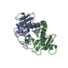
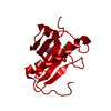
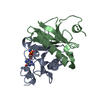
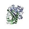
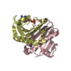
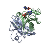
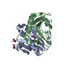
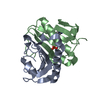
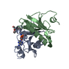
| Title | Human Histidine Triad Nucleotide Binding Protein 1 (hHint) IMP complex | ||||||
 Components Components | Histidine triad nucleotide-binding protein 1 | ||||||
 Keywords Keywords |  HYDROLASE / HINT / histidine triad / HIT HYDROLASE / HINT / histidine triad / HIT | ||||||
| Function / homology |  Function and homology information Function and homology informationpurine ribonucleotide catabolic process / adenylylsulfatase activity / Hydrolases; Acting on phosphorus-nitrogen bonds / adenosine 5'-monophosphoramidase activity / deSUMOylase activity / protein desumoylation / sulfur compound metabolic process / histone deacetylase complex / intrinsic apoptotic signaling pathway by p53 class mediator / positive regulation of calcium-mediated signaling ...purine ribonucleotide catabolic process / adenylylsulfatase activity / Hydrolases; Acting on phosphorus-nitrogen bonds / adenosine 5'-monophosphoramidase activity / deSUMOylase activity / protein desumoylation / sulfur compound metabolic process / histone deacetylase complex / intrinsic apoptotic signaling pathway by p53 class mediator / positive regulation of calcium-mediated signaling / protein kinase C binding / Hydrolases; Acting on peptide bonds (peptidases); Cysteine endopeptidases / cytoskeleton / hydrolase activity / nucleotide binding / regulation of DNA-templated transcription / signal transduction / proteolysis / extracellular exosome / nucleoplasm / nucleus / plasma membrane / cytosol / cytoplasmSimilarity search - Function | ||||||
| Biological species |  Homo sapiens (human) Homo sapiens (human) | ||||||
| Method | X-RAY DIFFRACTION / SYNCHROTRON / MOLECULAR REPLACEMENT / molecular replacement / Resolution: 1.533 Å | ||||||
 Authors Authors | Maize, K.M. / Finzel, B.C. | ||||||
| Funding support |  United States, 1items United States, 1items
| ||||||
 Citation Citation | Journal: Mol. Pharm. / Year: 2017 Title: A Crystal Structure Based Guide to the Design of Human Histidine Triad Nucleotide Binding Protein 1 (hHint1) Activated ProTides. Authors: Maize, K.M. / Shah, R. / Strom, A. / Kumarapperuma, S. / Zhou, A. / Wagner, C.R. / Finzel, B.C. | ||||||
| History |
|
- Structure visualization
| Structure viewer | Molecule: MolmilJmol/JSmol |
|---|
- Downloads & links
-Download
| PDBx/mmCIF format | 5km0.cif.gz | 117.5 KB | Display | PDBx/mmCIF format |
|---|---|---|---|---|
| PDB format | pdb5km0.ent.gz | 89.1 KB | Display | PDB format |
| PDBx/mmJSON format | 5km0.json.gz | Tree view | PDBx/mmJSON format | |
| Others |  Other downloads Other downloads |
-Validation report
| Arichive directory | https://data.pdbj.org/pub/pdb/validation_reports/km/5km0ftp://data.pdbj.org/pub/pdb/validation_reports/km/5km0 | HTTPS FTP |
|---|
-Related structure data
| Related structure data |  5klyC 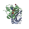 5klzC 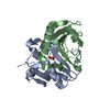 5km1C  5km2C  5km3C  5km4C  5km5C  5km6C  5km8C  5km9C  5kmaC  5kmbC  5wa8C  5wa9C  6b42C  1kpcS C: citing same article ( S: Starting model for refinement |
|---|---|
| Similar structure data |


-Links
 PDBj
PDBj


- Assembly
| Deposited unit | 
| ||||||||
|---|---|---|---|---|---|---|---|---|---|
| 1 | 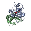
| ||||||||
| 2 | 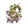
| ||||||||
| Unit cell |
|
-Components
| #1: Protein | Mass: 14096.188 Da / Num. of mol.: 4 Source method: isolated from a genetically manipulated source Source: (gene. exp.) Homo sapiens (human) / Gene: HINT1, HINT / Plasmid: pMCSG7 / Production host:  Escherichia coli (E. coli) / Strain (production host): Rosetta 2 pLysS / References: UniProt: P49773, Hydrolases Escherichia coli (E. coli) / Strain (production host): Rosetta 2 pLysS / References: UniProt: P49773, Hydrolases#2: Chemical | Inosinic acid  Mass: 348.206 Da / Num. of mol.: 2 / Source method: obtained synthetically / Formula: C10H13N4O8P Mass: 348.206 Da / Num. of mol.: 2 / Source method: obtained synthetically / Formula: C10H13N4O8P#3: Water | ChemComp-HOH / | Water Mass: 18.015 Da / Num. of mol.: 641 / Source method: isolated from a natural source / Formula: H2O Mass: 18.015 Da / Num. of mol.: 641 / Source method: isolated from a natural source / Formula: H2O |
|---|
-Experimental details
-Experiment
| Experiment | Method: X-RAY DIFFRACTION / Number of used crystals: 1 |
|---|
- Sample preparation
| Crystal | Density Matthews: 2.37 Å3/Da / Density % sol: 48.1 % |
|---|---|
| Crystal grow | Temperature: 293 K / Method: vapor diffusion, hanging drop / pH: 6.7 / Details: 100 mM MES, 34% PEG 8000 |
-Data collection
| Diffraction | Mean temperature: 100 K | |||||||||||||||
|---|---|---|---|---|---|---|---|---|---|---|---|---|---|---|---|---|
| Diffraction source | Source: SYNCHROTRON / Site: APS / Beamline: 17-ID / Wavelength: 1 Å | |||||||||||||||
| Detector | Type: DECTRIS PILATUS 6M / Detector: PIXEL / Date: Dec 15, 2013 | |||||||||||||||
| Radiation | Protocol: SINGLE WAVELENGTH / Monochromatic (M) / Laue (L): M / Scattering type: x-ray | |||||||||||||||
| Radiation wavelength | Wavelength: 1 Å / Relative weight: 1 | |||||||||||||||
| Reflection | Resolution: 1.533→64.325 Å / Num. obs: 77524 / % possible obs: 98.5 % / Redundancy: 3.2 % / Biso Wilson estimate: 12.33 Å2 / Net I/σ(I): 9.2 | |||||||||||||||
| Reflection shell |
|
-Phasing
| Phasing | Method: molecular replacement |
|---|
- Processing
| Software |
| |||||||||||||||||||||||||||||||||||||||||||||||||||||||||||||||||||||||||||||||||||||||||||||||||||||||||||||||||||||||||||||||||||||||||||||||||||||||||||||||||||||||||||||||||||||||||||||||||||||||||||
|---|---|---|---|---|---|---|---|---|---|---|---|---|---|---|---|---|---|---|---|---|---|---|---|---|---|---|---|---|---|---|---|---|---|---|---|---|---|---|---|---|---|---|---|---|---|---|---|---|---|---|---|---|---|---|---|---|---|---|---|---|---|---|---|---|---|---|---|---|---|---|---|---|---|---|---|---|---|---|---|---|---|---|---|---|---|---|---|---|---|---|---|---|---|---|---|---|---|---|---|---|---|---|---|---|---|---|---|---|---|---|---|---|---|---|---|---|---|---|---|---|---|---|---|---|---|---|---|---|---|---|---|---|---|---|---|---|---|---|---|---|---|---|---|---|---|---|---|---|---|---|---|---|---|---|---|---|---|---|---|---|---|---|---|---|---|---|---|---|---|---|---|---|---|---|---|---|---|---|---|---|---|---|---|---|---|---|---|---|---|---|---|---|---|---|---|---|---|---|---|---|---|---|---|---|
| Refinement | Method to determine structure: MOLECULAR REPLACEMENT Starting model: 1kpc Resolution: 1.533→41.182 Å / SU ML: 0.17 / Cross valid method: FREE R-VALUE / σ(F): 1.4 / Phase error: 24.03
| |||||||||||||||||||||||||||||||||||||||||||||||||||||||||||||||||||||||||||||||||||||||||||||||||||||||||||||||||||||||||||||||||||||||||||||||||||||||||||||||||||||||||||||||||||||||||||||||||||||||||||
| Solvent computation | Shrinkage radii: 0.9 Å / VDW probe radii: 1.11 Å | |||||||||||||||||||||||||||||||||||||||||||||||||||||||||||||||||||||||||||||||||||||||||||||||||||||||||||||||||||||||||||||||||||||||||||||||||||||||||||||||||||||||||||||||||||||||||||||||||||||||||||
| Displacement parameters | Biso max: 57.28 Å2 / Biso mean: 17.7118 Å2 / Biso min: 7.1 Å2 | |||||||||||||||||||||||||||||||||||||||||||||||||||||||||||||||||||||||||||||||||||||||||||||||||||||||||||||||||||||||||||||||||||||||||||||||||||||||||||||||||||||||||||||||||||||||||||||||||||||||||||
| Refinement step | Cycle: final / Resolution: 1.533→41.182 Å
| |||||||||||||||||||||||||||||||||||||||||||||||||||||||||||||||||||||||||||||||||||||||||||||||||||||||||||||||||||||||||||||||||||||||||||||||||||||||||||||||||||||||||||||||||||||||||||||||||||||||||||
| Refine LS restraints |
| |||||||||||||||||||||||||||||||||||||||||||||||||||||||||||||||||||||||||||||||||||||||||||||||||||||||||||||||||||||||||||||||||||||||||||||||||||||||||||||||||||||||||||||||||||||||||||||||||||||||||||
| LS refinement shell | Refine-ID: X-RAY DIFFRACTION / Total num. of bins used: 28
|
