 Movie
Movie Controller

[English] 日本語
 Yorodumi
- PDB-6eaf: CRYSTAL STRUCTURE OF HUMAN RESPIRATORY SYNCYTIAL VIRUS FUSION GLY... -
+ Open data

- Basic information
| Entry | Database: PDB / ID: 6eaf | |||||||||
|---|---|---|---|---|---|---|---|---|---|---|
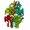
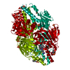
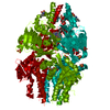
| Title | CRYSTAL STRUCTURE OF HUMAN RESPIRATORY SYNCYTIAL VIRUS FUSION GLYCOPROTEIN INHIBITOR ESCAPE VARIANT G143S STABILIZED IN THE PREFUSION STATE | |||||||||
 Components Components | Fusion glycoprotein F0 | |||||||||
 Keywords Keywords |  VIRAL PROTEIN / CLASS I VIRAL FUSION PROTEIN / FUSION / RESPIRATORY SYNCYTIAL VIRUS / PREFUSION / FUSION INHIBITOR VIRAL PROTEIN / CLASS I VIRAL FUSION PROTEIN / FUSION / RESPIRATORY SYNCYTIAL VIRUS / PREFUSION / FUSION INHIBITOR | |||||||||
| Function / homology |  Function and homology information Function and homology informationpositive regulation of syncytium formation by virus / host cell Golgi membrane / entry receptor-mediated virion attachment to host cell / symbiont entry into host cell / fusion of virus membrane with host plasma membrane / viral envelope / host cell plasma membrane / virion membrane / membrane / identical protein binding / plasma membraneSimilarity search - Function | |||||||||
| Biological species |  Human respiratory syncytial virus Human respiratory syncytial virus | |||||||||
| Method | X-RAY DIFFRACTION / SYNCHROTRON / MOLECULAR REPLACEMENT / Resolution: 3 Å | |||||||||
 Authors Authors | Battles, M.B. / McLellan, J.S. | |||||||||
| Funding support |  United States, 2items United States, 2items
| |||||||||
 Citation Citation | Journal: To be published Title: Structural Basis for Respiratory Syncytial Virus Fusion Inhibitor Resistance Authors: Battles, M.B. / McLellan, J.S. | |||||||||
| History |
|
- Structure visualization
| Structure viewer | Molecule: MolmilJmol/JSmol |
|---|
- Downloads & links
-Download
| PDBx/mmCIF format | 6eaf.cif.gz | 272 KB | Display | PDBx/mmCIF format |
|---|---|---|---|---|
| PDB format | pdb6eaf.ent.gz | 215.1 KB | Display | PDB format |
| PDBx/mmJSON format | 6eaf.json.gz | Tree view | PDBx/mmJSON format | |
| Others |  Other downloads Other downloads |
-Validation report
| Arichive directory | https://data.pdbj.org/pub/pdb/validation_reports/ea/6eafftp://data.pdbj.org/pub/pdb/validation_reports/ea/6eaf | HTTPS FTP |
|---|
-Related structure data
| Related structure data |  6eadC  6eaeC 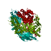 6eagC  6eahC  6eaiC  6eajC  6eakC 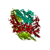 6ealC  6eamC  6eanC 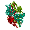 5ea4S S: Starting model for refinement C: citing same article ( |
|---|---|
| Similar structure data |


-Links
 PDBj
PDBj

- Assembly
| Deposited unit | 
| |||||||||||||||||||||||||||||||||||||||||||||||||||||||||||||||||||||||||||
|---|---|---|---|---|---|---|---|---|---|---|---|---|---|---|---|---|---|---|---|---|---|---|---|---|---|---|---|---|---|---|---|---|---|---|---|---|---|---|---|---|---|---|---|---|---|---|---|---|---|---|---|---|---|---|---|---|---|---|---|---|---|---|---|---|---|---|---|---|---|---|---|---|---|---|---|---|
| 1 |
| |||||||||||||||||||||||||||||||||||||||||||||||||||||||||||||||||||||||||||
| Unit cell |
| |||||||||||||||||||||||||||||||||||||||||||||||||||||||||||||||||||||||||||
| Noncrystallographic symmetry (NCS) | NCS domain:
NCS domain segments: Ens-ID: 1
|