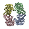
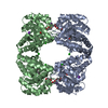
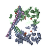
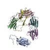
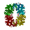
A: VACUOLAR PROTEIN SORTING-ASSOCIATED PROTEIN 75 B: VACUOLAR PROTEIN SORTING-ASSOCIATED PROTEIN 75 C: VACUOLAR PROTEIN SORTING-ASSOCIATED PROTEIN 75 D: VACUOLAR PROTEIN SORTING-ASSOCIATED PROTEIN 75
Resolution: 4→50.06 Å / Cor.coef. Fo:Fc: 0.928 / Cor.coef. Fo:Fc free: 0.915 / SU B: 63.669 / SU ML: 0.829 / Cross valid method: THROUGHOUT / ESU R Free: 0.93 / Stereochemistry target values: MAXIMUM LIKELIHOOD / Details: HYDROGENS HAVE BEEN ADDED IN THE RIDING POSITIONS.
Rfactor
Num. reflection
% reflection
Selection details
Rfree
0.26463
495
4.8 %
RANDOM
Rwork
0.22934
-
-
-
obs
0.23104
9771
98.88 %
-
Solvent computation
Ion probe radii: 0.8 Å / Shrinkage radii: 0.8 Å / VDW probe radii: 1.2 Å / Solvent model: MASK
 Movie
Movie Controller


 Open data
 Basic information
 Components
 Vacuole
  Keywords
 Function and homology information
 Authors
 Citation
 Structure visualization
 Downloads & links
 Other downloads

 PDBj
PDBj
 Assembly


 Sample preparation
 / Beamline: I04 / Wavelength: 0.97949
  Processing