 Movie
Movie Controller


+ Open data

- Basic information
| Entry | Database: PDB / ID: 4js8 | ||||||
|---|---|---|---|---|---|---|---|
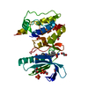
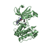
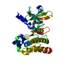
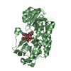
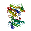
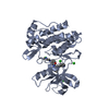
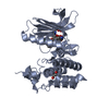
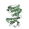
| Title | Crystal structure of TTK kinase domain with an inhibitor: 401348 | ||||||
 Components Components | Dual specificity protein kinase TTK | ||||||
 Keywords Keywords | Transferase/Transferase Inhibitor / Transferase-Transferase Inhibitor complex | ||||||
| Function / homology |  Function and homology information Function and homology informationprotein localization to meiotic spindle midzone / meiotic spindle assembly checkpoint signaling /  kinetochore binding / female meiosis chromosome segregation / protein localization to kinetochore / dual-specificity kinase / spindle organization / mitotic spindle assembly checkpoint signaling / protein serine/threonine/tyrosine kinase activity / mitotic spindle organization ...protein localization to meiotic spindle midzone / meiotic spindle assembly checkpoint signaling / kinetochore binding / female meiosis chromosome segregation / protein localization to kinetochore / dual-specificity kinase / spindle organization / mitotic spindle assembly checkpoint signaling / protein serine/threonine/tyrosine kinase activity / mitotic spindle organization / chromosome segregation / kinetochore / spindle / protein tyrosine kinase activity / phosphorylation / protein serine kinase activity / protein serine/threonine kinase activity / positive regulation of cell population proliferation / ATP binding / membrane / identical protein binding / nucleus / cytoplasm kinetochore binding / female meiosis chromosome segregation / protein localization to kinetochore / dual-specificity kinase / spindle organization / mitotic spindle assembly checkpoint signaling / protein serine/threonine/tyrosine kinase activity / mitotic spindle organization ...protein localization to meiotic spindle midzone / meiotic spindle assembly checkpoint signaling / kinetochore binding / female meiosis chromosome segregation / protein localization to kinetochore / dual-specificity kinase / spindle organization / mitotic spindle assembly checkpoint signaling / protein serine/threonine/tyrosine kinase activity / mitotic spindle organization / chromosome segregation / kinetochore / spindle / protein tyrosine kinase activity / phosphorylation / protein serine kinase activity / protein serine/threonine kinase activity / positive regulation of cell population proliferation / ATP binding / membrane / identical protein binding / nucleus / cytoplasmSimilarity search - Function | ||||||
| Biological species |  Homo sapiens (human) Homo sapiens (human) | ||||||
| Method | X-RAY DIFFRACTION / SYNCHROTRON / MOLECULAR REPLACEMENT / Resolution: 1.94 Å | ||||||
 Authors Authors | Qiu, W. / Plotnikov, A.N. / Plotnikova, O. / Feher, M. / Awrey, D.E. / Chirgadze, N.Y. | ||||||
 Citation Citation | Journal: To be Published Title: Crystal structure of TTK kinase domain with an inhibitor: 401348 Authors: Qiu, W. / Plotnikov, A.N. / Plotnikova, O. / Feher, M. / Awrey, D.E. / Chirgadze, N.Y. | ||||||
| History |
|
- Structure visualization
| Structure viewer | Molecule: MolmilJmol/JSmol |
|---|
- Downloads & links
-Download
| PDBx/mmCIF format | 4js8.cif.gz | 66.1 KB | Display | PDBx/mmCIF format |
|---|---|---|---|---|
| PDB format | pdb4js8.ent.gz | 52.5 KB | Display | PDB format |
| PDBx/mmJSON format | 4js8.json.gz | Tree view | PDBx/mmJSON format | |
| Others |  Other downloads Other downloads |
-Validation report
| Arichive directory | https://data.pdbj.org/pub/pdb/validation_reports/js/4js8ftp://data.pdbj.org/pub/pdb/validation_reports/js/4js8 | HTTPS FTP |
|---|
-Related structure data
| Related structure data | |
|---|---|
| Similar structure data |


-Links
 PDBj
PDBj- Assembly
| Deposited unit | 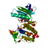
| ||||||||
|---|---|---|---|---|---|---|---|---|---|
| 1 |
| ||||||||
| 2 | 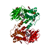
| ||||||||
| 3 | 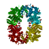
| ||||||||
| Unit cell |
|
-Components
-Protein , 1 types, 1 molecules A
| #1: Protein | Mass: 32619.260 Da / Num. of mol.: 1 / Fragment: kinase domain Source method: isolated from a genetically manipulated source Source: (gene. exp.) Homo sapiens (human) / Gene: TTK, MPS1, MPS1L1 / Production host:  Escherichia coli (E. coli) / References: UniProt: P33981, dual-specificity kinase Escherichia coli (E. coli) / References: UniProt: P33981, dual-specificity kinase |
|---|
-Non-polymers , 5 types, 80 molecules 


| #2: Chemical | ChemComp-1PF /  Mass: 419.559 Da / Num. of mol.: 1 / Source method: obtained synthetically / Formula: C26H33N3O2 Mass: 419.559 Da / Num. of mol.: 1 / Source method: obtained synthetically / Formula: C26H33N3O2 | ||||||
|---|---|---|---|---|---|---|---|
| #3: Chemical | Sulfate Mass: 96.063 Da / Num. of mol.: 2 / Source method: obtained synthetically / Formula: SO4 Mass: 96.063 Da / Num. of mol.: 2 / Source method: obtained synthetically / Formula: SO4#4: Chemical | Diethylene glycol Mass: 106.120 Da / Num. of mol.: 2 / Source method: obtained synthetically / Formula: C4H10O3 Mass: 106.120 Da / Num. of mol.: 2 / Source method: obtained synthetically / Formula: C4H10O3#5: Chemical | Glycerol Mass: 92.094 Da / Num. of mol.: 2 / Source method: obtained synthetically / Formula: C3H8O3 Mass: 92.094 Da / Num. of mol.: 2 / Source method: obtained synthetically / Formula: C3H8O3#6: Water | ChemComp-HOH / | WaterMass: 18.015 Da / Num. of mol.: 73 / Source method: isolated from a natural source / Formula: H2O |
-Experimental details
-Experiment
| Experiment | Method: X-RAY DIFFRACTION / Number of used crystals: 1 |
|---|
- Sample preparation
| Crystal | Density Matthews: 3.31 Å3/Da / Density % sol: 62.86 % |
|---|---|
| Crystal grow | Temperature: 293 K / Method: vapor diffusion, hanging drop / pH: 7.5 Details: 0.2M Ammonium Sulfate 0.1M HEPES pH7.5 25% PEG3350, VAPOR DIFFUSION, HANGING DROP, temperature 293K |
-Data collection
| Diffraction | Mean temperature: 100 K |
|---|---|
| Diffraction source | Source: SYNCHROTRON / Site: APS  / Beamline: 17-ID / Wavelength: 1 Å / Beamline: 17-ID / Wavelength: 1 Å |
| Detector | Type: PSI PILATUS 6M / Detector: PIXEL / Date: Jun 27, 2011 |
| Radiation | Monochromator: Si 111 CHANNEL / Protocol: SINGLE WAVELENGTH / Monochromatic (M) / Laue (L): M / Scattering type: x-ray |
| Radiation wavelength | Wavelength: 1 Å / Relative weight: 1 |
| Reflection | Resolution: 1.94→50 Å / Num. all: 32110 / Num. obs: 32110 / % possible obs: 99.5 % / Biso Wilson estimate: 49.7 Å2 |
| Reflection shell | Resolution: 1.94→2.05 Å / % possible all: 97 |
- Processing
| Software |
| ||||||||||||||||||||||||||||||||||||||||||||||||||||||||||||||||||||||||||||||||||||||||||||||||||||||||||||||||||
|---|---|---|---|---|---|---|---|---|---|---|---|---|---|---|---|---|---|---|---|---|---|---|---|---|---|---|---|---|---|---|---|---|---|---|---|---|---|---|---|---|---|---|---|---|---|---|---|---|---|---|---|---|---|---|---|---|---|---|---|---|---|---|---|---|---|---|---|---|---|---|---|---|---|---|---|---|---|---|---|---|---|---|---|---|---|---|---|---|---|---|---|---|---|---|---|---|---|---|---|---|---|---|---|---|---|---|---|---|---|---|---|---|---|---|---|
| Refinement | Method to determine structure: MOLECULAR REPLACEMENT / Resolution: 1.94→39.06 Å / Cor.coef. Fo:Fc: 0.938 / Cor.coef. Fo:Fc free: 0.9272 / SU R Cruickshank DPI: 0.141 / Cross valid method: THROUGHOUT / σ(F): 0 / Stereochemistry target values: Engh & Huber
| ||||||||||||||||||||||||||||||||||||||||||||||||||||||||||||||||||||||||||||||||||||||||||||||||||||||||||||||||||
| Displacement parameters | Biso mean: 63.95 Å2
| ||||||||||||||||||||||||||||||||||||||||||||||||||||||||||||||||||||||||||||||||||||||||||||||||||||||||||||||||||
| Refine analyze | Luzzati coordinate error obs: 0.339 Å | ||||||||||||||||||||||||||||||||||||||||||||||||||||||||||||||||||||||||||||||||||||||||||||||||||||||||||||||||||
| Refinement step | Cycle: LAST / Resolution: 1.94→39.06 Å
| ||||||||||||||||||||||||||||||||||||||||||||||||||||||||||||||||||||||||||||||||||||||||||||||||||||||||||||||||||
| Refine LS restraints |
| ||||||||||||||||||||||||||||||||||||||||||||||||||||||||||||||||||||||||||||||||||||||||||||||||||||||||||||||||||
| LS refinement shell | Resolution: 1.94→2 Å / Total num. of bins used: 16
|
