 Movie
Movie Controller

[English] 日本語
 Yorodumi
- PDB-6eak: CRYSTAL STRUCTURE OF FUSION INHIBITOR JNJ-2408068 IN COMPLEX WITH... -
+ Open data

- Basic information
| Entry | Database: PDB / ID: 6eak | |||||||||
|---|---|---|---|---|---|---|---|---|---|---|
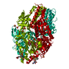
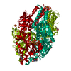
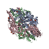
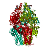
| Title | CRYSTAL STRUCTURE OF FUSION INHIBITOR JNJ-2408068 IN COMPLEX WITH HUMAN RESPIRATORY SYNCYTIAL VIRUS FUSION GLYCOPROTEIN ESCAPE VARIANT T400A STABILIZED IN THE PREFUSION STATE | |||||||||
 Components Components | Fusion glycoprotein F0 | |||||||||
 Keywords Keywords |  VIRAL PROTEIN / CLASS I VIRAL FUSION PROTEIN / FUSION / RESPIRATORY SYNCYTIAL VIRUS / PREFUSION / FUSION INHIBITOR VIRAL PROTEIN / CLASS I VIRAL FUSION PROTEIN / FUSION / RESPIRATORY SYNCYTIAL VIRUS / PREFUSION / FUSION INHIBITOR | |||||||||
| Function / homology |  Function and homology information Function and homology informationpositive regulation of syncytium formation by virus / host cell Golgi membrane / entry receptor-mediated virion attachment to host cell / symbiont entry into host cell / fusion of virus membrane with host plasma membrane / viral envelope / host cell plasma membrane / virion membrane / membrane / identical protein binding / plasma membraneSimilarity search - Function | |||||||||
| Biological species |  Human respiratory syncytial virus Human respiratory syncytial virus | |||||||||
| Method | X-RAY DIFFRACTION / SYNCHROTRON / MOLECULAR REPLACEMENT / Resolution: 2.6 Å | |||||||||
 Authors Authors | Battles, M.B. / McLellan, J.S. | |||||||||
| Funding support |  United States, 2items United States, 2items
| |||||||||
 Citation Citation | Journal: To be published Title: Structural Basis for Respiratory Syncytial Virus Fusion Inhibitor Resistance Authors: Battles, M.B. / McLellan, J.S. | |||||||||
| History |
|
- Structure visualization
| Structure viewer | Molecule: MolmilJmol/JSmol |
|---|
- Downloads & links
-Download
| PDBx/mmCIF format | 6eak.cif.gz | 108.3 KB | Display | PDBx/mmCIF format |
|---|---|---|---|---|
| PDB format | pdb6eak.ent.gz | 78.5 KB | Display | PDB format |
| PDBx/mmJSON format | 6eak.json.gz | Tree view | PDBx/mmJSON format | |
| Others |  Other downloads Other downloads |
-Validation report
| Arichive directory | https://data.pdbj.org/pub/pdb/validation_reports/ea/6eakftp://data.pdbj.org/pub/pdb/validation_reports/ea/6eak | HTTPS FTP |
|---|
-Related structure data
| Related structure data |  6eadC  6eaeC  6eafC  6eagC  6eahC  6eaiC  6eajC  6ealC  6eamC  6eanC  5ea4S S: Starting model for refinement C: citing same article ( |
|---|---|
| Similar structure data |

-Links
 PDBj
PDBj

- Assembly
| Deposited unit | 
| |||||||||||||||
|---|---|---|---|---|---|---|---|---|---|---|---|---|---|---|---|---|
| 1 | 
| |||||||||||||||
| Unit cell |
| |||||||||||||||
| Components on special symmetry positions |
|
-Components
-Protein , 1 types, 1 molecules F
| #1: Protein | Mass: 63188.320 Da / Num. of mol.: 1 / Fragment: RSV F ectodomain / Mutation: S155C, S290C, S190F, V207L, T400A Source method: isolated from a genetically manipulated source Source: (gene. exp.) Human respiratory syncytial virus / Plasmid: P(ALPHA)H / Production host:  Homo sapiens (human) / Strain (production host): HEK293 FREESTYLE / References: UniProt: W8RJF9, UniProt: P03420*PLUS Homo sapiens (human) / Strain (production host): HEK293 FREESTYLE / References: UniProt: W8RJF9, UniProt: P03420*PLUS |
|---|
-Non-polymers , 5 types, 64 molecules 


| #2: Chemical | ChemComp-SO4 / Sulfate Mass: 96.063 Da / Num. of mol.: 6 / Source method: obtained synthetically / Formula: SO4 Mass: 96.063 Da / Num. of mol.: 6 / Source method: obtained synthetically / Formula: SO4#3: Chemical | ChemComp-NHE / | CHES (buffer) Mass: 207.290 Da / Num. of mol.: 1 / Source method: obtained synthetically / Formula: C8H17NO3S / Comment: pH buffer*YM Mass: 207.290 Da / Num. of mol.: 1 / Source method: obtained synthetically / Formula: C8H17NO3S / Comment: pH buffer*YM#4: Chemical | ChemComp-NI / | Nickel Mass: 58.693 Da / Num. of mol.: 1 / Source method: obtained synthetically / Formula: Ni Mass: 58.693 Da / Num. of mol.: 1 / Source method: obtained synthetically / Formula: Ni#5: Chemical | ChemComp-5NK / |  Mass: 394.513 Da / Num. of mol.: 1 / Source method: obtained synthetically / Formula: C22H30N6O / Feature type: SUBJECT OF INVESTIGATION Mass: 394.513 Da / Num. of mol.: 1 / Source method: obtained synthetically / Formula: C22H30N6O / Feature type: SUBJECT OF INVESTIGATION#6: Water | ChemComp-HOH / | WaterMass: 18.015 Da / Num. of mol.: 55 / Source method: isolated from a natural source / Formula: H2O |
|---|
-Experimental details
-Experiment
| Experiment | Method: X-RAY DIFFRACTION / Number of used crystals: 1 |
|---|
- Sample preparation
| Crystal | Density Matthews: 3.15 Å3/Da / Density % sol: 60.98 % / Mosaicity: 0.66 ° |
|---|---|
| Crystal grow | Temperature: 293 K / Method: vapor diffusion, hanging drop / pH: 9.5 / Details: 1.48M K/Na tartrate, 0.2M LiSO4, 0.1M CHES pH 9.5 |
-Data collection
| Diffraction | Mean temperature: 80 K |
|---|---|
| Diffraction source | Source: SYNCHROTRON / Site: APS / Beamline: 19-ID / Wavelength: 0.9793 Å |
| Detector | Type: ADSC QUANTUM 315 / Detector: CCD / Date: Nov 8, 2016 |
| Radiation | Monochromator: Si(111) / Protocol: SINGLE WAVELENGTH / Monochromatic (M) / Laue (L): M / Scattering type: x-ray |
| Radiation wavelength | Wavelength: 0.9793 Å / Relative weight: 1 |
| Reflection | Resolution: 2.6→59.56 Å / Num. obs: 25761 / % possible obs: 100 % / Redundancy: 14.6 % / Biso Wilson estimate: 55.15 Å2 / CC1/2: 0.999 / Rmerge(I) obs: 0.13 / Rpim(I) all: 0.035 / Rrim(I) all: 0.135 / Net I/σ(I): 15.5 / Num. measured all: 376488 / Scaling rejects: 9 |
| Reflection shell | Resolution: 2.6→2.72 Å / Redundancy: 15.6 % / Rmerge(I) obs: 1.267 / Num. unique obs: 3084 / CC1/2: 0.795 / Rpim(I) all: 0.33 / Rrim(I) all: 1.31 / % possible all: 100 |
- Processing
| Software |
| ||||||||||||||||||||||||||||||||||||||||||||||||||||||||||||
|---|---|---|---|---|---|---|---|---|---|---|---|---|---|---|---|---|---|---|---|---|---|---|---|---|---|---|---|---|---|---|---|---|---|---|---|---|---|---|---|---|---|---|---|---|---|---|---|---|---|---|---|---|---|---|---|---|---|---|---|---|---|
| Refinement | Method to determine structure: MOLECULAR REPLACEMENT Starting model: 5EA4 Resolution: 2.6→59.556 Å / SU ML: 0.35 / Cross valid method: THROUGHOUT / σ(F): 1.35 / Phase error: 22.01
| ||||||||||||||||||||||||||||||||||||||||||||||||||||||||||||
| Solvent computation | Shrinkage radii: 0.9 Å / VDW probe radii: 1.11 Å | ||||||||||||||||||||||||||||||||||||||||||||||||||||||||||||
| Displacement parameters | Biso max: 155.93 Å2 / Biso mean: 62.1724 Å2 / Biso min: 23.73 Å2 | ||||||||||||||||||||||||||||||||||||||||||||||||||||||||||||
| Refinement step | Cycle: final / Resolution: 2.6→59.556 Å
| ||||||||||||||||||||||||||||||||||||||||||||||||||||||||||||
| LS refinement shell | Refine-ID: X-RAY DIFFRACTION / Rfactor Rfree error: 0 / Total num. of bins used: 9 / % reflection obs: 100 %
|
