 Movie
Movie Controller

[English] 日本語
 Yorodumi
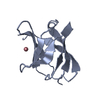
- PDB-6cd2: Crystal structure of the PapC usher bound to the chaperone-adhesi... -
+ Open data

- Basic information
| Entry | Database: PDB / ID: 6cd2 | ||||||||||||
|---|---|---|---|---|---|---|---|---|---|---|---|---|---|
| Title | Crystal structure of the PapC usher bound to the chaperone-adhesin PapD-PapG | ||||||||||||
 Components Components |
| ||||||||||||
 Keywords Keywords |  MEMBRANE PROTEIN/CHAPERONE / outer membrane usher / P pilus assembly / usher activation / MEMBRANE PROTEIN / MEMBRANE PROTEIN-CHAPERONE complex MEMBRANE PROTEIN/CHAPERONE / outer membrane usher / P pilus assembly / usher activation / MEMBRANE PROTEIN / MEMBRANE PROTEIN-CHAPERONE complex | ||||||||||||
| Function / homology |  Function and homology information Function and homology informationfimbrial usher porin activity / pilus assembly / pilus / chaperone-mediated protein folding / cell wall organization / cell outer membrane / outer membrane-bounded periplasmic space / carbohydrate binding / cell adhesion / extracellular region / identical protein bindingSimilarity search - Function | ||||||||||||
| Biological species |  Escherichia coli (E. coli) Escherichia coli (E. coli) | ||||||||||||
| Method | X-RAY DIFFRACTION / SYNCHROTRON / MOLECULAR REPLACEMENT / Resolution: 3.7 Å | ||||||||||||
 Authors Authors | Omattage, N.S. / Deng, Z. / Yuan, P. / Hultgren, S.J. | ||||||||||||
| Funding support |  United States, 3items United States, 3items
| ||||||||||||
 Citation Citation | Journal: Nat Microbiol / Year: 2018 Title: Structural basis for usher activation and intramolecular subunit transfer in P pilus biogenesis in Escherichia coli. Authors: Omattage, N.S. / Deng, Z. / Pinkner, J.S. / Dodson, K.W. / Almqvist, F. / Yuan, P. / Hultgren, S.J. | ||||||||||||
| History |
|
- Structure visualization
| Structure viewer | Molecule: MolmilJmol/JSmol |
|---|
- Downloads & links
-Download
| PDBx/mmCIF format | 6cd2.cif.gz | 462.6 KB | Display | PDBx/mmCIF format |
|---|---|---|---|---|
| PDB format | pdb6cd2.ent.gz | 380.5 KB | Display | PDB format |
| PDBx/mmJSON format | 6cd2.json.gz | Tree view | PDBx/mmJSON format | |
| Others |  Other downloads Other downloads |
-Validation report
| Arichive directory | https://data.pdbj.org/pub/pdb/validation_reports/cd/6cd2ftp://data.pdbj.org/pub/pdb/validation_reports/cd/6cd2 | HTTPS FTP |
|---|
-Related structure data


-Links
 PDBj
PDBj- Assembly
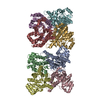
| Deposited unit | 
| ||||||||
|---|---|---|---|---|---|---|---|---|---|
| 1 |
| ||||||||
| Unit cell |
|
-Components
| #1: Protein | Mass: 25031.303 Da / Num. of mol.: 1 Source method: isolated from a genetically manipulated source Source: (gene. exp.) Escherichia coli (E. coli) / Gene: papD / Production host: Escherichia coli (E. coli) / References: UniProt: P15319 |
|---|---|
| #2: Protein | Mass: 35519.094 Da / Num. of mol.: 1 Source method: isolated from a genetically manipulated source Source: (gene. exp.) Escherichia coli (E. coli) / Production host: Escherichia coli (E. coli) / References: UniProt: Q47450 |
| #3: Protein | Mass: 80472.062 Da / Num. of mol.: 1 Source method: isolated from a genetically manipulated source Source: (gene. exp.) Escherichia coli (E. coli) / Gene: PapC / Production host: Escherichia coli (E. coli) / References: UniProt: P07110*PLUS |
| Sequence details | The poly-UNK fragments in PapC sequence represent portions that the residue identities cannot be ...The poly-UNK fragments in PapC sequence represent portions that the residue identities cannot be determined from electron density. The full sequence of the entity is VEFNTDVLDA |
-Experimental details
-Experiment
| Experiment | Method: X-RAY DIFFRACTION / Number of used crystals: 1 |
|---|
- Sample preparation
| Crystal | Density Matthews: 5.12 Å3/Da / Density % sol: 75.99 % |
|---|---|
| Crystal grow | Temperature: 293 K / Method: vapor diffusion, sitting drop Details: 50 mM sodium citrate pH 5.0-6.0, 50 mM lithium sulfate, 50 mM sodium sulfate and 7-14% PEG 4000 |
-Data collection
| Diffraction | Mean temperature: 100 K |
|---|---|
| Diffraction source | Source: SYNCHROTRON / Site: APS / Beamline: 24-ID-E / Wavelength: 0.979 Å |
| Detector | Type: DECTRIS EIGER X 16M / Detector: PIXEL / Date: Jun 7, 2017 |
| Radiation | Protocol: SINGLE WAVELENGTH / Monochromatic (M) / Laue (L): M / Scattering type: x-ray |
| Radiation wavelength | Wavelength: 0.979 Å / Relative weight: 1 |
| Reflection | Resolution: 3.7→50 Å / Num. obs: 27152 / % possible obs: 98.5 % / Redundancy: 6 % / Rmerge(I) obs: 0.122 / Rpim(I) all: 0.054 / Rrim(I) all: 0.134 / Net I/σ(I): 15.9 |
| Reflection shell | Resolution: 3.7→3.83 Å / Redundancy: 5.1 % / Mean I/σ(I) obs: 1.5 / Num. unique obs: 3048 / Rpim(I) all: 0.448 / % possible all: 92.5 |
- Processing
| Software |
| ||||||||||||||||||||||||||||||||||||||||||||||||||||||||||||||||||||||||||||||||||||||||||||||||||||||||||||||||||||||||||||||||||||||||||||||||||||||||||||||||||||||||||||||||||||||
|---|---|---|---|---|---|---|---|---|---|---|---|---|---|---|---|---|---|---|---|---|---|---|---|---|---|---|---|---|---|---|---|---|---|---|---|---|---|---|---|---|---|---|---|---|---|---|---|---|---|---|---|---|---|---|---|---|---|---|---|---|---|---|---|---|---|---|---|---|---|---|---|---|---|---|---|---|---|---|---|---|---|---|---|---|---|---|---|---|---|---|---|---|---|---|---|---|---|---|---|---|---|---|---|---|---|---|---|---|---|---|---|---|---|---|---|---|---|---|---|---|---|---|---|---|---|---|---|---|---|---|---|---|---|---|---|---|---|---|---|---|---|---|---|---|---|---|---|---|---|---|---|---|---|---|---|---|---|---|---|---|---|---|---|---|---|---|---|---|---|---|---|---|---|---|---|---|---|---|---|---|---|---|---|
| Refinement | Method to determine structure: MOLECULAR REPLACEMENT Starting model: 2VQI, 2WMP, 1J8S, 3L48 Resolution: 3.7→50.01 Å / Cor.coef. Fo:Fc: 0.832 / Cor.coef. Fo:Fc free: 0.791 / SU B: 91.949 / SU ML: 0.615 / Cross valid method: THROUGHOUT / ESU R Free: 0.77 / Stereochemistry target values: MAXIMUM LIKELIHOOD / Details: HYDROGENS HAVE BEEN ADDED IN THE RIDING POSITIONS
| ||||||||||||||||||||||||||||||||||||||||||||||||||||||||||||||||||||||||||||||||||||||||||||||||||||||||||||||||||||||||||||||||||||||||||||||||||||||||||||||||||||||||||||||||||||||
| Solvent computation | Ion probe radii: 0.8 Å / Shrinkage radii: 0.8 Å / VDW probe radii: 1.2 Å / Solvent model: MASK | ||||||||||||||||||||||||||||||||||||||||||||||||||||||||||||||||||||||||||||||||||||||||||||||||||||||||||||||||||||||||||||||||||||||||||||||||||||||||||||||||||||||||||||||||||||||
| Displacement parameters | Biso mean: 135.172 Å2
| ||||||||||||||||||||||||||||||||||||||||||||||||||||||||||||||||||||||||||||||||||||||||||||||||||||||||||||||||||||||||||||||||||||||||||||||||||||||||||||||||||||||||||||||||||||||
| Refinement step | Cycle: 1 / Resolution: 3.7→50.01 Å
| ||||||||||||||||||||||||||||||||||||||||||||||||||||||||||||||||||||||||||||||||||||||||||||||||||||||||||||||||||||||||||||||||||||||||||||||||||||||||||||||||||||||||||||||||||||||
| Refine LS restraints |
|