 Movie
Movie Controller

[English] 日本語
 Yorodumi
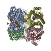
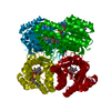
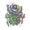
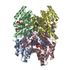
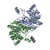
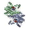
- PDB-1zmo: Apo structure of haloalcohol dehalogenase HheA of Arthrobacter sp. AD2 -
+ Open data

- Basic information
| Entry | Database: PDB / ID: 1zmo | ||||||
|---|---|---|---|---|---|---|---|
| Title | Apo structure of haloalcohol dehalogenase HheA of Arthrobacter sp. AD2 | ||||||
 Components Components | halohydrin dehalogenase | ||||||
 Keywords Keywords | LYASE / halohydrin dehalogenase / haloalcohol dehalogenase / short-chain dehydrogenase/reductase family | ||||||
| Function / homology | Enoyl-(Acyl carrier protein) reductase / Short-chain dehydrogenase/reductase SDR / NAD(P)-binding Rossmann-like Domain / NAD(P)-binding domain superfamily / Rossmann fold / 3-Layer(aba) Sandwich / Alpha Beta / Halohydrin dehalogenase Function and homology information Function and homology information | ||||||
| Biological species |  Arthrobacter sp. AD2 (bacteria) Arthrobacter sp. AD2 (bacteria) | ||||||
| Method | X-RAY DIFFRACTION / SYNCHROTRON / MOLECULAR REPLACEMENT / Resolution: 2 Å | ||||||
 Authors Authors | de Jong, R.M. / Kalk, K.H. / Tang, L. / Janssen, D.B. / Dijkstra, B.W. | ||||||
 Citation Citation | Journal: J.Bacteriol. / Year: 2006 Title: The X-ray structure of the haloalcohol dehalogenase HheA from Arthrobacter sp. strain AD2: insight into enantioselectivity and halide binding in the haloalcohol dehalogenase family. Authors: de Jong, R.M. / Kalk, K.H. / Tang, L. / Janssen, D.B. / Dijkstra, B.W. | ||||||
| History |
|
- Structure visualization
| Structure viewer | Molecule: MolmilJmol/JSmol |
|---|
- Downloads & links
-Download
| PDBx/mmCIF format | 1zmo.cif.gz | 374.4 KB | Display | PDBx/mmCIF format |
|---|---|---|---|---|
| PDB format | pdb1zmo.ent.gz | 305.9 KB | Display | PDB format |
| PDBx/mmJSON format | 1zmo.json.gz | Tree view | PDBx/mmJSON format | |
| Others |  Other downloads Other downloads |
-Validation report
| Arichive directory | https://data.pdbj.org/pub/pdb/validation_reports/zm/1zmoftp://data.pdbj.org/pub/pdb/validation_reports/zm/1zmo | HTTPS FTP |
|---|
-Related structure data
| Related structure data | 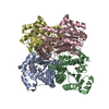 1pwxS S: Starting model for refinement |
|---|---|
| Similar structure data |


-Links
 PDBj
PDBj

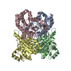
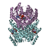
- Assembly
| Deposited unit | 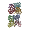
| ||||||||
|---|---|---|---|---|---|---|---|---|---|
| 1 | 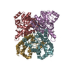
| ||||||||
| 2 | 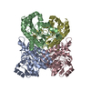
| ||||||||
| Unit cell |
| ||||||||
| Details | There are two biologically active tetramers in the asymmetric unit. |
-Components
| #1: Protein | Mass: 26451.004 Da / Num. of mol.: 8 Source method: isolated from a genetically manipulated source Source: (gene. exp.) Arthrobacter sp. AD2 (bacteria) / Species (production host): Escherichia coli / Production host: Escherichia coli BL21(DE3) (bacteria) / Strain (production host): BL21(DE3) / References: UniProt: Q93MS3, EC: 4.5.-.-#2: Water | ChemComp-HOH / | Water Mass: 18.015 Da / Num. of mol.: 862 / Source method: isolated from a natural source / Formula: H2O Mass: 18.015 Da / Num. of mol.: 862 / Source method: isolated from a natural source / Formula: H2O |
|---|
-Experimental details
-Experiment
| Experiment | Method: X-RAY DIFFRACTION / Number of used crystals: 1 |
|---|
- Sample preparation
| Crystal | Density Matthews: 2.52 Å3/Da / Density % sol: 50.8 % |
|---|---|
| Crystal grow | Temperature: 298 K / Method: vapor diffusion, hanging drop / pH: 6.5 Details: 22% w/v PEG 8000, 100 mM MES buffer, 100 mM Magnesium Acetate, 50 mM Sodium Formate, pH 6.5, VAPOR DIFFUSION, HANGING DROP, temperature 298K |
-Data collection
| Diffraction | Mean temperature: 100 K |
|---|---|
| Diffraction source | Source: SYNCHROTRON / Site: ESRF  / Beamline: ID14-2 / Wavelength: 0.93 Å / Beamline: ID14-2 / Wavelength: 0.93 Å |
| Detector | Type: ADSC QUANTUM 4 / Detector: CCD / Date: Apr 9, 2003 |
| Radiation | Monochromator: Si / Protocol: SINGLE WAVELENGTH / Monochromatic (M) / Laue (L): M / Scattering type: x-ray |
| Radiation wavelength | Wavelength: 0.93 Å / Relative weight: 1 |
| Reflection | Resolution: 2→25 Å / Num. obs: 120343 / % possible obs: 89.4 % / Observed criterion σ(F): 2 / Observed criterion σ(I): 2 / Biso Wilson estimate: 10.2 Å2 |
| Reflection shell | Resolution: 2→2.09 Å / % possible all: 89.4 |
- Processing
| Software |
| ||||||||||||||||||||||||||||||||||||
|---|---|---|---|---|---|---|---|---|---|---|---|---|---|---|---|---|---|---|---|---|---|---|---|---|---|---|---|---|---|---|---|---|---|---|---|---|---|
| Refinement | Method to determine structure: MOLECULAR REPLACEMENT Starting model: PDB entry 1PWX Resolution: 2→20.05 Å / Rfactor Rfree error: 0.004 / Data cutoff high absF: 1374501.74 / Data cutoff high rms absF: 1374501.74 / Data cutoff low absF: 0 / Isotropic thermal model: RESTRAINED / Cross valid method: THROUGHOUT / σ(F): 0 / Stereochemistry target values: Engh & Huber
| ||||||||||||||||||||||||||||||||||||
| Solvent computation | Solvent model: FLAT MODEL / Bsol: 64.2387 Å2 / ksol: 0.416166 e/Å3 | ||||||||||||||||||||||||||||||||||||
| Displacement parameters | Biso mean: 20.1 Å2
| ||||||||||||||||||||||||||||||||||||
| Refine analyze |
| ||||||||||||||||||||||||||||||||||||
| Refinement step | Cycle: LAST / Resolution: 2→20.05 Å
| ||||||||||||||||||||||||||||||||||||
| Refine LS restraints |
| ||||||||||||||||||||||||||||||||||||
| LS refinement shell | Resolution: 2→2.13 Å / Rfactor Rfree error: 0.011 / Total num. of bins used: 6
| ||||||||||||||||||||||||||||||||||||
| Xplor file |
|
