 Movie
Movie Controller

[English] 日本語
 Yorodumi
- PDB-1mxh: Crystal Structure of Substrate Complex of Putative Pteridine Redu... -
+ Open data

- Basic information
| Entry | Database: PDB / ID: 1mxh | ||||||
|---|---|---|---|---|---|---|---|
| Title | Crystal Structure of Substrate Complex of Putative Pteridine Reductase 2 (PTR2) from Trypanosoma cruzi | ||||||
 Components Components | PTERIDINE REDUCTASE 2 | ||||||
 Keywords Keywords | OXIDOREDUCTASE / SDR TOPOLOGY / PROTEIN-SUBSTRATE COMPLEX | ||||||
| Function / homology |  Function and homology information Function and homology information | ||||||
| Biological species |  Trypanosoma cruzi (eukaryote) Trypanosoma cruzi (eukaryote) | ||||||
| Method | X-RAY DIFFRACTION / SYNCHROTRON / MOLECULAR REPLACEMENT / Resolution: 2.2 Å | ||||||
 Authors Authors | Schormann, N. / Pal, B. / Senkovich, O. / Carson, M. / Howard, A. / Smith, C. / Delucas, L. / Chattopadhyay, D. | ||||||
 Citation Citation | Journal: J.Struct.Biol. / Year: 2005 Title: Crystal structure of Trypanosoma cruzi pteridine reductase 2 in complex with a substrate and an inhibitor. Authors: Schormann, N. / Pal, B. / Senkovich, O. / Carson, M. / Howard, A. / Smith, C. / Delucas, L. / Chattopadhyay, D. #1: Journal: Acta Crystallogr.,Sect.D / Year: 2001Title: Expression, purification, crystallization and preliminary crystallographic analysis of recombinant pteridine reductase of Trypanosoma cruzi Authors: Schormann, N. / Pal, B. / Chattopadhyay, D. | ||||||
| History |
| ||||||
| Remark 600 | Heterogen Ligand coordinates were taken from PDB entry 1RF7. The coordinate file was missing a ...Heterogen Ligand coordinates were taken from PDB entry 1RF7. The coordinate file was missing a carboxylate group in the glutamate portion. No correction was made since the glutamate portion of ligand DHF shows no clear density in this structure. |
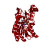
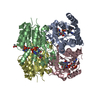
- Structure visualization
| Structure viewer | Molecule: MolmilJmol/JSmol |
|---|
- Downloads & links
-Download
| PDBx/mmCIF format | 1mxh.cif.gz | 209.8 KB | Display | PDBx/mmCIF format |
|---|---|---|---|---|
| PDB format | pdb1mxh.ent.gz | 169 KB | Display | PDB format |
| PDBx/mmJSON format | 1mxh.json.gz | Tree view | PDBx/mmJSON format | |
| Others |  Other downloads Other downloads |
-Validation report
| Arichive directory | https://data.pdbj.org/pub/pdb/validation_reports/mx/1mxhftp://data.pdbj.org/pub/pdb/validation_reports/mx/1mxh | HTTPS FTP |
|---|
-Related structure data
| Related structure data |  1mxfSC S: Starting model for refinement C: citing same article ( |
|---|---|
| Similar structure data |


-Links
 PDBj
PDBj


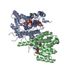
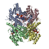
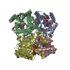
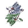
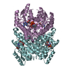
- Assembly
| Deposited unit | 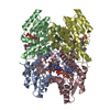
| ||||||||
|---|---|---|---|---|---|---|---|---|---|
| 1 |
| ||||||||
| Unit cell |
|
-Components
| #1: Protein | Mass: 29181.277 Da / Num. of mol.: 4 Source method: isolated from a genetically manipulated source Source: (gene. exp.) Trypanosoma cruzi (eukaryote) / Strain: Sylvio, X10 / Gene: ptr2 / Plasmid: pET15b / Production host:  Escherichia coli (E. coli) / Strain (production host): BL21(DE3)pLysS / References: UniProt: Q8I814 Escherichia coli (E. coli) / Strain (production host): BL21(DE3)pLysS / References: UniProt: Q8I814#2: Chemical | ChemComp-NAP / Nicotinamide adenine dinucleotide phosphate  Mass: 743.405 Da / Num. of mol.: 4 / Source method: obtained synthetically / Formula: C21H28N7O17P3 Mass: 743.405 Da / Num. of mol.: 4 / Source method: obtained synthetically / Formula: C21H28N7O17P3#3: Chemical | ChemComp-DHF / Dihydrofolic acid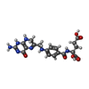  Mass: 443.413 Da / Num. of mol.: 4 / Source method: obtained synthetically / Formula: C19H21N7O6 Mass: 443.413 Da / Num. of mol.: 4 / Source method: obtained synthetically / Formula: C19H21N7O6#4: Water | ChemComp-HOH / | Water Mass: 18.015 Da / Num. of mol.: 457 / Source method: isolated from a natural source / Formula: H2O Mass: 18.015 Da / Num. of mol.: 457 / Source method: isolated from a natural source / Formula: H2O |
|---|
-Experimental details
-Experiment
| Experiment | Method: X-RAY DIFFRACTION / Number of used crystals: 1 |
|---|
- Sample preparation
| Crystal | Density Matthews: 2.3 Å3/Da / Density % sol: 46.7 % | |||||||||||||||||||||||||||||||||||||||||||||||||
|---|---|---|---|---|---|---|---|---|---|---|---|---|---|---|---|---|---|---|---|---|---|---|---|---|---|---|---|---|---|---|---|---|---|---|---|---|---|---|---|---|---|---|---|---|---|---|---|---|---|---|
| Crystal grow | Temperature: 293 K / pH: 6.5 Details: Sodium acetate, cacodylate buffer, pH 6.50, temperature 293K | |||||||||||||||||||||||||||||||||||||||||||||||||
| Crystal grow | *PLUS Temperature: 293 K / pH: 8 / Method: vapor diffusion, hanging dropDetails: Schormann, N., (2001) Acta Crystallogr.,Sect.D, 57, 1671. | |||||||||||||||||||||||||||||||||||||||||||||||||
| Components of the solutions | *PLUS
|
-Data collection
| Diffraction | Mean temperature: 103 K |
|---|---|
| Diffraction source | Source: SYNCHROTRON / Site: APS  / Beamline: 17-ID / Wavelength: 0.9537 / Beamline: 17-ID / Wavelength: 0.9537 |
| Detector | Type: MARRESEARCH / Detector: CCD / Date: Dec 20, 2000 / Details: MIRRORS |
| Radiation | Monochromator: GRAPHITE / Protocol: SINGLE WAVELENGTH / Monochromatic (M) / Laue (L): M / Scattering type: x-ray |
| Radiation wavelength | Wavelength: 0.9537 Å / Relative weight: 1 |
| Reflection | Resolution: 1.77→36 Å / Num. obs: 87817 / % possible obs: 79.8 % / Observed criterion σ(I): 0 / Redundancy: 1.9 % / Biso Wilson estimate: 17.7 Å2 / Rmerge(I) obs: 0.025 / Net I/σ(I): 12.4 |
| Reflection | *PLUS Highest resolution: 2.3 Å / Lowest resolution: 50 Å / Num. obs: 47858 / % possible obs: 95.7 % / Redundancy: 2.4 % / Rmerge(I) obs: 0.059 |
| Reflection shell | *PLUS Highest resolution: 2.3 Å / Lowest resolution: 2.44 Å / % possible obs: 91.2 % / Redundancy: 1.8 % / Num. unique obs: 7590 / Rmerge(I) obs: 0.182 / Mean I/σ(I) obs: 3.9 |
- Processing
| Software |
| ||||||||||||||||||||||||||||||||||||||||||||||||||||||||||||||||||||||||||||||||
|---|---|---|---|---|---|---|---|---|---|---|---|---|---|---|---|---|---|---|---|---|---|---|---|---|---|---|---|---|---|---|---|---|---|---|---|---|---|---|---|---|---|---|---|---|---|---|---|---|---|---|---|---|---|---|---|---|---|---|---|---|---|---|---|---|---|---|---|---|---|---|---|---|---|---|---|---|---|---|---|---|---|
| Refinement | Method to determine structure: MOLECULAR REPLACEMENT Starting model: INHIBITOR COMPLEX SOLVED BY MAD METHOD (PDBID 1MXF) Resolution: 2.2→19.45 Å / Rfactor Rfree error: 0.005 / Data cutoff high absF: 220622 / Data cutoff high rms absF: 220622 / Data cutoff low absF: 0 / Isotropic thermal model: RESTRAINED / Cross valid method: THROUGHOUT / σ(F): 1 / Stereochemistry target values: ENGH & HUBER
| ||||||||||||||||||||||||||||||||||||||||||||||||||||||||||||||||||||||||||||||||
| Solvent computation | Solvent model: FLAT MODEL / Bsol: 52.225 Å2 / ksol: 0.361 e/Å3 | ||||||||||||||||||||||||||||||||||||||||||||||||||||||||||||||||||||||||||||||||
| Displacement parameters | Biso mean: 31.1 Å2
| ||||||||||||||||||||||||||||||||||||||||||||||||||||||||||||||||||||||||||||||||
| Refine analyze |
| ||||||||||||||||||||||||||||||||||||||||||||||||||||||||||||||||||||||||||||||||
| Refinement step | Cycle: LAST / Resolution: 2.2→19.45 Å
| ||||||||||||||||||||||||||||||||||||||||||||||||||||||||||||||||||||||||||||||||
| Refine LS restraints |
| ||||||||||||||||||||||||||||||||||||||||||||||||||||||||||||||||||||||||||||||||
| LS refinement shell | Resolution: 2.2→2.34 Å / Rfactor Rfree error: 0.014 / Total num. of bins used: 6
| ||||||||||||||||||||||||||||||||||||||||||||||||||||||||||||||||||||||||||||||||
| Xplor file |
| ||||||||||||||||||||||||||||||||||||||||||||||||||||||||||||||||||||||||||||||||
| Refinement | *PLUS Highest resolution: 2.3 Å / Lowest resolution: 50 Å / Rfactor Rfree: 0.252 / Rfactor Rwork: 0.2 | ||||||||||||||||||||||||||||||||||||||||||||||||||||||||||||||||||||||||||||||||
| Solvent computation | *PLUS | ||||||||||||||||||||||||||||||||||||||||||||||||||||||||||||||||||||||||||||||||
| Displacement parameters | *PLUS | ||||||||||||||||||||||||||||||||||||||||||||||||||||||||||||||||||||||||||||||||
| Refine LS restraints | *PLUS
| ||||||||||||||||||||||||||||||||||||||||||||||||||||||||||||||||||||||||||||||||
| LS refinement shell | *PLUS Highest resolution: 2.3 Å / Lowest resolution: 2.44 Å / Rfactor Rfree: 0.288 / Rfactor Rwork: 0.246 |
