 Movie
Movie Controller

[English] 日本語
 Yorodumi
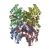
- PDB-6zzq: Crystal structure of (R)-3-hydroxybutyrate dehydrogenase from Aci... -
+ Open data

- Basic information
| Entry | Database: PDB / ID: 6zzq | ||||||
|---|---|---|---|---|---|---|---|
| Title | Crystal structure of (R)-3-hydroxybutyrate dehydrogenase from Acinetobacter baumannii complexed with NAD+ and acetoacetate | ||||||
 Components Components | 3-hydroxybutyrate dehydrogenase | ||||||
 Keywords Keywords | OXIDOREDUCTASE / (R)-3-hydroxybutyrate dehydrogenase / short-chain dehydrogenase/reductase / mesophilic enzyme | ||||||
| Function / homology |  Function and homology information Function and homology information | ||||||
| Biological species |  Acinetobacter baumannii (bacteria) Acinetobacter baumannii (bacteria) | ||||||
| Method | X-RAY DIFFRACTION / SYNCHROTRON / MOLECULAR REPLACEMENT / Resolution: 1.93 Å | ||||||
 Authors Authors | Machado, T.F.G. / da Silva, R.G. / Gloster, T.M. / McMahon, S.A. / Oehler, V. | ||||||
| Funding support |  United Kingdom, 1items United Kingdom, 1items
| ||||||
 Citation Citation | Journal: Acs Catalysis / Year: 2020 Title: Dissecting the Mechanism of ( R )-3-Hydroxybutyrate Dehydrogenase by Kinetic Isotope Effects, Protein Crystallography, and Computational Chemistry. Authors: Machado, T.F.G. / Purg, M. / McMahon, S.A. / Read, B.J. / Oehler, V. / Aqvist, J. / Gloster, T.M. / da Silva, R.G. | ||||||
| History |
|
- Structure visualization
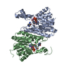
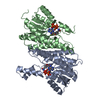
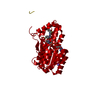
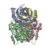
| Structure viewer | Molecule: MolmilJmol/JSmol |
|---|
- Downloads & links
-Download
| PDBx/mmCIF format | 6zzq.cif.gz | 66.6 KB | Display | PDBx/mmCIF format |
|---|---|---|---|---|
| PDB format | pdb6zzq.ent.gz | 47.4 KB | Display | PDB format |
| PDBx/mmJSON format | 6zzq.json.gz | Tree view | PDBx/mmJSON format | |
| Others |  Other downloads Other downloads |
-Validation report
| Arichive directory | https://data.pdbj.org/pub/pdb/validation_reports/zz/6zzqftp://data.pdbj.org/pub/pdb/validation_reports/zz/6zzq | HTTPS FTP |
|---|
-Related structure data
| Related structure data |  6zzoC 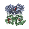 6zzpC 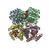 6zzsC 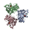 2v2hS S: Starting model for refinement C: citing same article ( |
|---|---|
| Similar structure data |

-Links
 PDBj
PDBj


- Assembly
| Deposited unit | 
| ||||||||||||
|---|---|---|---|---|---|---|---|---|---|---|---|---|---|
| 1 | 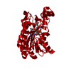
| ||||||||||||
| Unit cell |
| ||||||||||||
| Components on special symmetry positions |
|
-Components
| #1: Protein | Mass: 27567.816 Da / Num. of mol.: 1 Source method: isolated from a genetically manipulated source Source: (gene. exp.) Acinetobacter baumannii (bacteria) / Gene: A7M79_09600, BGC29_06470 / Production host: Escherichia coli BL21(DE3) (bacteria) / Variant (production host): Gold / References: UniProt: A0A1E3M3N6 | ||||
|---|---|---|---|---|---|
| #2: Chemical | ChemComp-NAD / Nicotinamide adenine dinucleotide  Mass: 663.425 Da / Num. of mol.: 1 / Source method: obtained synthetically / Formula: C21H27N7O14P2 / Feature type: SUBJECT OF INVESTIGATION / Comment: NAD*YM Mass: 663.425 Da / Num. of mol.: 1 / Source method: obtained synthetically / Formula: C21H27N7O14P2 / Feature type: SUBJECT OF INVESTIGATION / Comment: NAD*YM | ||||
| #3: Chemical | Acetoacetic acid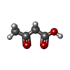  Mass: 102.089 Da / Num. of mol.: 2 / Source method: obtained synthetically / Formula: C4H6O3 / Feature type: SUBJECT OF INVESTIGATION Mass: 102.089 Da / Num. of mol.: 2 / Source method: obtained synthetically / Formula: C4H6O3 / Feature type: SUBJECT OF INVESTIGATION#4: Water | ChemComp-HOH / | Water Mass: 18.015 Da / Num. of mol.: 121 / Source method: isolated from a natural source / Formula: H2O Mass: 18.015 Da / Num. of mol.: 121 / Source method: isolated from a natural source / Formula: H2OHas ligand of interest | Y | |
-Experimental details
-Experiment
| Experiment | Method: X-RAY DIFFRACTION / Number of used crystals: 1 |
|---|
- Sample preparation
| Crystal | Density Matthews: 2.1 Å3/Da / Density % sol: 41.54 % |
|---|---|
| Crystal grow | Temperature: 298 K / Method: vapor diffusion, sitting drop / pH: 7 / Details: 15% (v/w) PEG 3350, 0.1 M succinic acid |
-Data collection
| Diffraction | Mean temperature: 175 K / Serial crystal experiment: N |
|---|---|
| Diffraction source | Source: SYNCHROTRON / Site: Diamond / Beamline: I04 / Wavelength: 0.9795 Å |
| Detector | Type: DECTRIS EIGER X 16M / Detector: PIXEL / Date: Sep 24, 2017 |
| Radiation | Protocol: SINGLE WAVELENGTH / Monochromatic (M) / Laue (L): M / Scattering type: x-ray |
| Radiation wavelength | Wavelength: 0.9795 Å / Relative weight: 1 |
| Reflection | Resolution: 1.93→53.57 Å / Num. obs: 18880 / % possible obs: 100 % / Redundancy: 24.5 % / CC1/2: 0.999 / Rmerge(I) obs: 0.141 / Rpim(I) all: 0.029 / Rrim(I) all: 0.144 / Net I/σ(I): 15.7 |
| Reflection shell | Resolution: 1.93→1.98 Å / Redundancy: 16.7 % / Rmerge(I) obs: 3.014 / Num. unique obs: 1344 / CC1/2: 0.827 / R split: 1.6 / Rpim(I) all: 0.759 / % possible all: 100 |
- Processing
| Software |
| ||||||||||||||||||||||||||||||||||||||||||||||||||||||||||||
|---|---|---|---|---|---|---|---|---|---|---|---|---|---|---|---|---|---|---|---|---|---|---|---|---|---|---|---|---|---|---|---|---|---|---|---|---|---|---|---|---|---|---|---|---|---|---|---|---|---|---|---|---|---|---|---|---|---|---|---|---|---|
| Refinement | Method to determine structure: MOLECULAR REPLACEMENT Starting model: 2V2H Resolution: 1.93→53.57 Å / Cor.coef. Fo:Fc: 0.964 / Cor.coef. Fo:Fc free: 0.942 / SU B: 6.279 / SU ML: 0.17 / Cross valid method: THROUGHOUT / σ(F): 0 / ESU R: 0.203 / ESU R Free: 0.185 / Stereochemistry target values: MAXIMUM LIKELIHOOD Details: HYDROGENS HAVE BEEN ADDED IN THE RIDING POSITIONS U VALUES : REFINED INDIVIDUALLY
| ||||||||||||||||||||||||||||||||||||||||||||||||||||||||||||
| Solvent computation | Ion probe radii: 0.8 Å / Shrinkage radii: 0.8 Å / VDW probe radii: 1.2 Å / Solvent model: MASK | ||||||||||||||||||||||||||||||||||||||||||||||||||||||||||||
| Displacement parameters | Biso max: 82.55 Å2 / Biso mean: 33.566 Å2 / Biso min: 16.18 Å2
| ||||||||||||||||||||||||||||||||||||||||||||||||||||||||||||
| Refinement step | Cycle: final / Resolution: 1.93→53.57 Å
| ||||||||||||||||||||||||||||||||||||||||||||||||||||||||||||
| Refine LS restraints |
| ||||||||||||||||||||||||||||||||||||||||||||||||||||||||||||
| LS refinement shell | Resolution: 1.931→1.981 Å / Rfactor Rfree error: 0 / Total num. of bins used: 20
|
