 Movie
Movie Controller

[English] 日本語
 Yorodumi
- PDB-4n5l: Crystal structure of (R)-3-hydroxybutyryl-CoA dehydrogenase from ... -
+ Open data

- Basic information
| Entry | Database: PDB / ID: 4n5l | ||||||
|---|---|---|---|---|---|---|---|
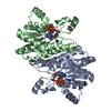
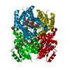
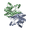
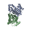
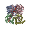
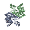
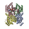
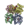
| Title | Crystal structure of (R)-3-hydroxybutyryl-CoA dehydrogenase from Ralstonia eutropha | ||||||
 Components Components | Acetoacetyl-CoA reductase | ||||||
 Keywords Keywords | OXIDOREDUCTASE / alpha/beta structure | ||||||
| Function / homology |  Function and homology informationacetoacetyl-CoA reductase / acetoacetyl-CoA reductase activity / poly-hydroxybutyrate biosynthetic process / cytoplasm Function and homology informationacetoacetyl-CoA reductase / acetoacetyl-CoA reductase activity / poly-hydroxybutyrate biosynthetic process / cytoplasmSimilarity search - Function | ||||||
| Biological species |  Ralstonia eutropha (bacteria) Ralstonia eutropha (bacteria) | ||||||
| Method | X-RAY DIFFRACTION / SYNCHROTRON / MOLECULAR REPLACEMENT / molecular replacement / Resolution: 1.65 Å | ||||||
 Authors Authors | Kim, J.-E. / Kim, S. / Kim, K.-J. | ||||||
 Citation Citation | Journal: Biochem.Biophys.Res.Commun. / Year: 2014 Title: Crystal structure of (R)-3-hydroxybutyryl-CoA dehydrogenase PhaB from Ralstonia eutropha Authors: Kim, J.-E. / Chang, J.H. / Kim, E.-J. / Kim, K.-J. | ||||||
| History |
|
- Structure visualization
| Structure viewer | Molecule: MolmilJmol/JSmol |
|---|
- Downloads & links
-Download
| PDBx/mmCIF format | 4n5l.cif.gz | 109.3 KB | Display | PDBx/mmCIF format |
|---|---|---|---|---|
| PDB format | pdb4n5l.ent.gz | 82.7 KB | Display | PDB format |
| PDBx/mmJSON format | 4n5l.json.gz | Tree view | PDBx/mmJSON format | |
| Others |  Other downloads Other downloads |
-Validation report
| Arichive directory | https://data.pdbj.org/pub/pdb/validation_reports/n5/4n5lftp://data.pdbj.org/pub/pdb/validation_reports/n5/4n5l | HTTPS FTP |
|---|
-Related structure data
| Related structure data |  4n5mC  4n5nC  1q7bS S: Starting model for refinement C: citing same article ( |
|---|---|
| Similar structure data |


-Links
 PDBj
PDBj


- Assembly
| Deposited unit | 
| ||||||||
|---|---|---|---|---|---|---|---|---|---|
| 1 | 
| ||||||||
| Unit cell |
|
-Components
| #1: Protein | / (R)-3-hydroxybutyrylCoA dehydrogenase Mass: 29399.158 Da / Num. of mol.: 2 Source method: isolated from a genetically manipulated source Source: (gene. exp.) Ralstonia eutropha (bacteria) / Strain: H16 / Gene: phbB / Plasmid: pProEX HTa / Production host: Escherichia coli (E. coli) / Strain (production host): B834 / References: UniProt: P14697, acetoacetyl-CoA reductase#2: Water | ChemComp-HOH / | Water Mass: 18.015 Da / Num. of mol.: 311 / Source method: isolated from a natural source / Formula: H2O Mass: 18.015 Da / Num. of mol.: 311 / Source method: isolated from a natural source / Formula: H2O |
|---|
-Experimental details
-Experiment
| Experiment | Method: X-RAY DIFFRACTION / Number of used crystals: 1 |
|---|
- Sample preparation
| Crystal | Density Matthews: 2.42 Å3/Da / Density % sol: 49.18 % |
|---|---|
| Crystal grow | Temperature: 295 K / Method: vapor diffusion, hanging drop / pH: 6.5 Details: ethanol,citrate, lithium sulfate, pH 6.5, vapor diffusion, hanging drop, temperature 295K |
-Data collection
| Diffraction | Mean temperature: 100 K | |||||||||||||||||||||||||||||||||||||||||||||||||||||||||||||||
|---|---|---|---|---|---|---|---|---|---|---|---|---|---|---|---|---|---|---|---|---|---|---|---|---|---|---|---|---|---|---|---|---|---|---|---|---|---|---|---|---|---|---|---|---|---|---|---|---|---|---|---|---|---|---|---|---|---|---|---|---|---|---|---|---|
| Diffraction source | Source: SYNCHROTRON / Site: PAL/PLS  / Beamline: 7A (6B, 6C1) / Wavelength: 0.979 Å / Beamline: 7A (6B, 6C1) / Wavelength: 0.979 Å | |||||||||||||||||||||||||||||||||||||||||||||||||||||||||||||||
| Detector | Type: ADSC QUANTUM 270 / Detector: CCD / Date: Dec 16, 2012 | |||||||||||||||||||||||||||||||||||||||||||||||||||||||||||||||
| Radiation | Monochromator: Double Crystal Monochromator / Protocol: SINGLE WAVELENGTH / Monochromatic (M) / Laue (L): M / Scattering type: x-ray | |||||||||||||||||||||||||||||||||||||||||||||||||||||||||||||||
| Radiation wavelength | Wavelength: 0.979 Å / Relative weight: 1 | |||||||||||||||||||||||||||||||||||||||||||||||||||||||||||||||
| Reflection | Resolution: 1.65→68.64 Å / Num. all: 68610 / Num. obs: 59854 / % possible obs: 87.2 % / Observed criterion σ(F): 2 / Observed criterion σ(I): 2 / Redundancy: 6.8 % / Rmerge(I) obs: 0.059 / Rsym value: 0.051 / Net I/σ(I): 30 | |||||||||||||||||||||||||||||||||||||||||||||||||||||||||||||||
| Reflection shell |
|
-Phasing
| Phasing | Method: molecular replacement |
|---|
- Processing
| Software |
| |||||||||||||||||||||||||||||||||||||||||||||||||||||||||||||||||||||||||||
|---|---|---|---|---|---|---|---|---|---|---|---|---|---|---|---|---|---|---|---|---|---|---|---|---|---|---|---|---|---|---|---|---|---|---|---|---|---|---|---|---|---|---|---|---|---|---|---|---|---|---|---|---|---|---|---|---|---|---|---|---|---|---|---|---|---|---|---|---|---|---|---|---|---|---|---|---|
| Refinement | Method to determine structure: MOLECULAR REPLACEMENT Starting model: 1Q7B Resolution: 1.65→32.18 Å / Cor.coef. Fo:Fc: 0.945 / Cor.coef. Fo:Fc free: 0.922 / Occupancy max: 1 / Occupancy min: 1 / SU B: 2.942 / SU ML: 0.097 / Cross valid method: THROUGHOUT / σ(F): 0 / ESU R: 0.135 / ESU R Free: 0.136 / Stereochemistry target values: MAXIMUM LIKELIHOOD Details: HYDROGENS HAVE BEEN ADDED IN THE RIDING POSITIONS U VALUES: REFINED INDIVIDUALLY
| |||||||||||||||||||||||||||||||||||||||||||||||||||||||||||||||||||||||||||
| Solvent computation | Ion probe radii: 0.8 Å / Shrinkage radii: 0.8 Å / VDW probe radii: 1.2 Å / Solvent model: MASK | |||||||||||||||||||||||||||||||||||||||||||||||||||||||||||||||||||||||||||
| Displacement parameters | Biso max: 76.54 Å2 / Biso mean: 23.1289 Å2 / Biso min: 8.55 Å2
| |||||||||||||||||||||||||||||||||||||||||||||||||||||||||||||||||||||||||||
| Refinement step | Cycle: LAST / Resolution: 1.65→32.18 Å
| |||||||||||||||||||||||||||||||||||||||||||||||||||||||||||||||||||||||||||
| Refine LS restraints |
| |||||||||||||||||||||||||||||||||||||||||||||||||||||||||||||||||||||||||||
| LS refinement shell | Resolution: 1.651→1.693 Å / Total num. of bins used: 20
|
