 Movie
Movie Controller

[English] 日本語
 Yorodumi
- PDB-1pwx: Crystal structure of the haloalcohol dehalogenase HheC complexed ... -
+ Open data

- Basic information
| Entry | Database: PDB / ID: 1pwx | ||||||
|---|---|---|---|---|---|---|---|
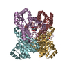
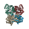
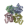
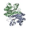
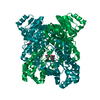
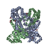
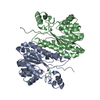
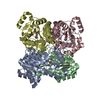
| Title | Crystal structure of the haloalcohol dehalogenase HheC complexed with bromide | ||||||
 Components Components | halohydrin dehalogenase | ||||||
 Keywords Keywords | LYASE / HALOALCOHOL DEHALOGENASE / HALOHYDRIN DEHALOGENASE / HALOHYDRIN HYDROGEN-HALIDE LYASE / SDR FAMILY / SHORT-CHAIN DEHYDROGENASE/REDUCTASE / ROSSMANN FOLD | ||||||
| Function / homology | Enoyl-(Acyl carrier protein) reductase / Short-chain dehydrogenase/reductase SDR / NAD(P)-binding Rossmann-like Domain / NAD(P)-binding domain superfamily / Rossmann fold / 3-Layer(aba) Sandwich / Alpha Beta / BROMIDE ION / Halohydrin dehalogenase Function and homology information Function and homology information | ||||||
| Biological species |  Agrobacterium tumefaciens (bacteria) Agrobacterium tumefaciens (bacteria) | ||||||
| Method | X-RAY DIFFRACTION / SYNCHROTRON / MOLECULAR REPLACEMENT / Resolution: 1.8 Å | ||||||
 Authors Authors | de Jong, R.M. / Tiesinga, J.J.W. / Rozeboom, H.J. / Kalk, K.H. / Tang, L. / Janssen, D.B. / Dijkstra, B.W. | ||||||
 Citation Citation | Journal: EMBO J. / Year: 2003 Title: Structure and Mechanism of a Bacterial Haloalcohol Dehalogenase: a new variation of the short-chain dehydrogenase/reductase fold without an NAD(P)H binding site Authors: de Jong, R.M. / Tiesinga, J.J.W. / Rozeboom, H.J. / Kalk, K.H. / Tang, L. / Janssen, D.B. / Dijkstra, B.W. | ||||||
| History |
|
- Structure visualization
| Structure viewer | Molecule: MolmilJmol/JSmol |
|---|
- Downloads & links
-Download
| PDBx/mmCIF format | 1pwx.cif.gz | 194.1 KB | Display | PDBx/mmCIF format |
|---|---|---|---|---|
| PDB format | pdb1pwx.ent.gz | 157.6 KB | Display | PDB format |
| PDBx/mmJSON format | 1pwx.json.gz | Tree view | PDBx/mmJSON format | |
| Others |  Other downloads Other downloads |
-Validation report
| Arichive directory | https://data.pdbj.org/pub/pdb/validation_reports/pw/1pwxftp://data.pdbj.org/pub/pdb/validation_reports/pw/1pwx | HTTPS FTP |
|---|
-Related structure data


-Links
 PDBj
PDBj


- Assembly
| Deposited unit | 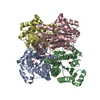
| ||||||||
|---|---|---|---|---|---|---|---|---|---|
| 1 |
| ||||||||
| Unit cell |
| ||||||||
| Details | The asymmetric unit contains one biological functional tetramer |
-Components
| #1: Protein | Mass: 27979.666 Da / Num. of mol.: 4 Source method: isolated from a genetically manipulated source Source: (gene. exp.) Agrobacterium tumefaciens (bacteria) / Plasmid: pGEF / Species (production host): Escherichia coli / Production host: Escherichia coli BL21(DE3) (bacteria) / Strain (production host): BL21(DE3) / References: UniProt: Q93D82#2: Chemical | ChemComp-BR / Bromide  Mass: 79.904 Da / Num. of mol.: 8 / Source method: obtained synthetically / Formula: Br Mass: 79.904 Da / Num. of mol.: 8 / Source method: obtained synthetically / Formula: Br#3: Water | ChemComp-HOH / | Water Mass: 18.015 Da / Num. of mol.: 525 / Source method: isolated from a natural source / Formula: H2O Mass: 18.015 Da / Num. of mol.: 525 / Source method: isolated from a natural source / Formula: H2O |
|---|
-Experimental details
-Experiment
| Experiment | Method: X-RAY DIFFRACTION / Number of used crystals: 1 |
|---|
- Sample preparation
| Crystal | Density Matthews: 2.79 Å3/Da / Density % sol: 55.95 % | ||||||||||||||||||
|---|---|---|---|---|---|---|---|---|---|---|---|---|---|---|---|---|---|---|---|
| Crystal grow | Temperature: 293 K / Method: vapor diffusion, hanging drop / pH: 6.9 Details: ammonium sulfate, bis-tris buffer, pH 6.9, VAPOR DIFFUSION, HANGING DROP, temperature 293K | ||||||||||||||||||
| Crystal grow | *PLUS Method: vapor diffusion, hanging drop | ||||||||||||||||||
| Components of the solutions | *PLUS
|
-Data collection
| Diffraction | Mean temperature: 100 K |
|---|---|
| Diffraction source | Source: SYNCHROTRON / Site: ELETTRA  / Beamline: 5.2R / Wavelength: 0.93 Å / Beamline: 5.2R / Wavelength: 0.93 Å |
| Detector | Type: MARRESEARCH / Detector: IMAGE PLATE / Date: Dec 13, 1999 |
| Radiation | Protocol: SINGLE WAVELENGTH / Monochromatic (M) / Laue (L): M / Scattering type: x-ray |
| Radiation wavelength | Wavelength: 0.93 Å / Relative weight: 1 |
| Reflection | Resolution: 1.8→35 Å / % possible obs: 95.2 % / Observed criterion σ(F): 2 / Observed criterion σ(I): 2 / Redundancy: 2.5 % / Biso Wilson estimate: 13.7 Å2 / Rmerge(I) obs: 0.079 / Rsym value: 0.066 / Net I/σ(I): 23.4 |
| Reflection shell | Resolution: 1.8→1.86 Å / Redundancy: 1.05 % / Rmerge(I) obs: 0.152 / Mean I/σ(I) obs: 3.2 / Num. unique all: 4234 / Rsym value: 0.164 / % possible all: 96.3 |
| Reflection | *PLUS Num. obs: 63725 / Num. measured all: 1561167 / Rmerge(I) obs: 0.066 |
| Reflection shell | *PLUS Highest resolution: 1.8 Å / Lowest resolution: 1.9 Å / % possible obs: 46.4 % / Rmerge(I) obs: 0.17 |
- Processing
| Software |
| |||||||||||||||||||||||||
|---|---|---|---|---|---|---|---|---|---|---|---|---|---|---|---|---|---|---|---|---|---|---|---|---|---|---|
| Refinement | Method to determine structure: MOLECULAR REPLACEMENT Starting model: Initial model from a low resolution MAD dataset, which has not been refined Resolution: 1.8→38.76 Å / Rfactor Rfree error: 0.004 / Data cutoff high absF: 2198381.76 / Data cutoff high rms absF: 2198381.76 / Data cutoff low absF: 0 / Isotropic thermal model: GROUP / Cross valid method: THROUGHOUT / σ(F): 2 / σ(I): 2 / Stereochemistry target values: Engh & Huber Details: CRYSTAL IS A PERFECT TWIN: R/R FREE ARE TWINNED-R/R FREE
| |||||||||||||||||||||||||
| Solvent computation | Solvent model: FLAT MODEL / Bsol: 52.699 Å2 / ksol: 0.383578 e/Å3 | |||||||||||||||||||||||||
| Displacement parameters | Biso mean: 16.8 Å2
| |||||||||||||||||||||||||
| Refine analyze |
| |||||||||||||||||||||||||
| Refinement step | Cycle: LAST / Resolution: 1.8→38.76 Å
| |||||||||||||||||||||||||
| Refine LS restraints |
| |||||||||||||||||||||||||
| LS refinement shell | Resolution: 1.8→1.86 Å / Rfactor Rfree error: 0.013 / Total num. of bins used: 10
| |||||||||||||||||||||||||
| Xplor file |
| |||||||||||||||||||||||||
| Software | *PLUS Name: CNS / Version: 1.1 / Classification: refinement | |||||||||||||||||||||||||
| Refinement | *PLUS Highest resolution: 1.82 Å / Lowest resolution: 35 Å / % reflection Rfree: 5 % / Rfactor Rfree: 0.309 / Rfactor Rwork: 0.233 | |||||||||||||||||||||||||
| Solvent computation | *PLUS | |||||||||||||||||||||||||
| Displacement parameters | *PLUS | |||||||||||||||||||||||||
| Refine LS restraints | *PLUS
|
