 Movie
Movie Controller

[English] 日本語
 Yorodumi
- PDB-1ye6: Crystal structure of the Lys-274 to Arg mutant of Candida tenuis ... -
+ Open data

- Basic information
| Entry | Database: PDB / ID: 1ye6 | ||||||
|---|---|---|---|---|---|---|---|
| Title | Crystal structure of the Lys-274 to Arg mutant of Candida tenuis xylose reductase (AKR2B5) bound to NADP+ | ||||||
 Components Components | NAD(P)H-dependent D-xylose reductase | ||||||
 Keywords Keywords |  OXIDOREDUCTASE / beta-alpha-barrel AKR aldo-keto reductase coenzyme specificity NADP OXIDOREDUCTASE / beta-alpha-barrel AKR aldo-keto reductase coenzyme specificity NADP | ||||||
| Function / homology |  Function and homology information Function and homology informationD-xylose reductase [NAD(P)H] / D-xylose reductase (NADPH) activity / D-xylose catabolic process Similarity search - Function | ||||||
| Biological species |  Candida tenuis (fungus) Candida tenuis (fungus) | ||||||
| Method | X-RAY DIFFRACTION / SYNCHROTRON / isomourphous / Resolution: 2.3 Å | ||||||
 Authors Authors | Leitgeb, S. / Petschacher, B. / Wilson, D.K. / Nidetzky, B. | ||||||
 Citation Citation | Journal: Febs Lett. / Year: 2005 Title: Fine tuning of coenzyme specificity in family 2 aldo-keto reductases revealed by crystal structures of the Lys-274-->Arg mutant of Candida tenuis xylose reductase (AKR2B5) bound to NAD(+) and NADP(+). Authors: Leitgeb, S. / Petschacher, B. / Wilson, D.K. / Nidetzky, B. | ||||||
| History |
|
- Structure visualization
| Structure viewer | Molecule: MolmilJmol/JSmol |
|---|
- Downloads & links
-Download
| PDBx/mmCIF format | 1ye6.cif.gz | 278 KB | Display | PDBx/mmCIF format |
|---|---|---|---|---|
| PDB format | pdb1ye6.ent.gz | 224.7 KB | Display | PDB format |
| PDBx/mmJSON format | 1ye6.json.gz | Tree view | PDBx/mmJSON format | |
| Others |  Other downloads Other downloads |
-Validation report
| Arichive directory | https://data.pdbj.org/pub/pdb/validation_reports/ye/1ye6ftp://data.pdbj.org/pub/pdb/validation_reports/ye/1ye6 | HTTPS FTP |
|---|
-Related structure data
| Related structure data |  1ye4C 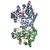 1k8cS C: citing same article ( S: Starting model for refinement |
|---|---|
| Similar structure data |


-Links
 PDBj
PDBj
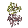
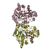
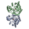
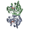
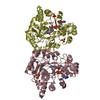
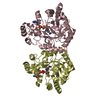
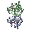
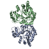
- Assembly
| Deposited unit | 
| ||||||||||||
|---|---|---|---|---|---|---|---|---|---|---|---|---|---|
| 1 | 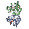
| ||||||||||||
| 2 | 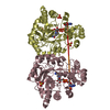
| ||||||||||||
| 3 | 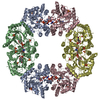
| ||||||||||||
| 4 | 
| ||||||||||||
| 5 | 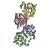
| ||||||||||||
| Unit cell |
| ||||||||||||
| Components on special symmetry positions |
|
-Components
| #1: Protein | Mass: 36090.215 Da / Num. of mol.: 4 / Mutation: K274R Source method: isolated from a genetically manipulated source Source: (gene. exp.) Candida tenuis (fungus) / Gene: XYL1, XYLR / Plasmid: pET11 / Species (production host): Escherichia coli / Production host:  Escherichia coli BL21(DE3) (bacteria) / Strain (production host): BL 21(DE3) Escherichia coli BL21(DE3) (bacteria) / Strain (production host): BL 21(DE3)References: UniProt: O74237, Oxidoreductases; Acting on the CH-OH group of donors; With NAD+ or NADP+ as acceptor#2: Chemical | ChemComp-SO4 / Sulfate  Mass: 96.063 Da / Num. of mol.: 8 / Source method: obtained synthetically / Formula: SO4 Mass: 96.063 Da / Num. of mol.: 8 / Source method: obtained synthetically / Formula: SO4#3: Chemical | Nicotinamide adenine dinucleotide phosphate  Mass: 743.405 Da / Num. of mol.: 2 / Source method: obtained synthetically / Formula: C21H28N7O17P3 Mass: 743.405 Da / Num. of mol.: 2 / Source method: obtained synthetically / Formula: C21H28N7O17P3#4: Chemical | Nicotinamide adenine dinucleotide  Mass: 663.425 Da / Num. of mol.: 2 / Source method: obtained synthetically / Formula: C21H27N7O14P2 / Comment: NAD*YM Mass: 663.425 Da / Num. of mol.: 2 / Source method: obtained synthetically / Formula: C21H27N7O14P2 / Comment: NAD*YM#5: Water | ChemComp-HOH / | Water Mass: 18.015 Da / Num. of mol.: 760 / Source method: isolated from a natural source / Formula: H2O Mass: 18.015 Da / Num. of mol.: 760 / Source method: isolated from a natural source / Formula: H2O |
|---|
-Experimental details
-Experiment
| Experiment | Method: X-RAY DIFFRACTION / Number of used crystals: 1 |
|---|
- Sample preparation
| Crystal | Density Matthews: 3.19 Å3/Da / Density % sol: 61.5 % |
|---|---|
| Crystal grow | Temperature: 298 K / Method: vapor diffusion, hanging drop / pH: 6.2 Details: Ammonium sulfate, sodium citrate, sodium acetate, pH 6.2, VAPOR DIFFUSION, HANGING DROP, temperature 298K |
-Data collection
| Diffraction | Mean temperature: 100 K |
|---|---|
| Diffraction source | Source: SYNCHROTRON / Site: SSRL  / Beamline: BL9-1 / Wavelength: 0.953695 Å / Beamline: BL9-1 / Wavelength: 0.953695 Å |
| Detector | Type: MARRESEARCH / Detector: IMAGE PLATE / Date: Jan 1, 2004 |
| Radiation | Monochromator: single crystal Si(311) bent monochromator / Protocol: SINGLE WAVELENGTH / Monochromatic (M) / Laue (L): M / Scattering type: x-ray |
| Radiation wavelength | Wavelength: 0.953695 Å / Relative weight: 1 |
| Reflection | Resolution: 2.3→30 Å / Num. all: 80204 / Num. obs: 80204 / % possible obs: 99.7 % / Observed criterion σ(I): 0 / Rmerge(I) obs: 0.094 / Net I/σ(I): 13.38 |
| Reflection shell | Resolution: 2.3→2.38 Å / Rmerge(I) obs: 0.347 / Mean I/σ(I) obs: 4.47 / % possible all: 98.8 |
- Processing
| Software |
| |||||||||||||||||||||||||
|---|---|---|---|---|---|---|---|---|---|---|---|---|---|---|---|---|---|---|---|---|---|---|---|---|---|---|
| Refinement | Method to determine structure: isomourphous Starting model: PDB-entry 1K8C Resolution: 2.3→30 Å / σ(F): 0
| |||||||||||||||||||||||||
| Refinement step | Cycle: LAST / Resolution: 2.3→30 Å
| |||||||||||||||||||||||||
| Refine LS restraints |
|
