 Movie
Movie Controller

[English] 日本語
 Yorodumi
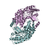
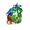
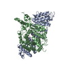
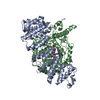
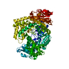
- PDB-3lg0: Structure of Plasmodium falciparum ornithine delta-aminotransferase -
+ Open data

- Basic information
| Entry | Database: PDB / ID: 3lg0 | ||||||
|---|---|---|---|---|---|---|---|
| Title | Structure of Plasmodium falciparum ornithine delta-aminotransferase | ||||||
 Components Components | Ornithine aminotransferase | ||||||
 Keywords Keywords | TRANSFERASE / Activation of Plasmodium falciparum ornithine delta-aminotransferase / delta-aminotransferase / pyridoxal-5-phosphate-dependent enzyme / ornithine / alpha-ketoglutarate / Plasmodium falciparum / Aminotransferase / Pyridoxal phosphate | ||||||
| Function / homology |  Function and homology information: / ornithine aminotransferase activity / ornithine aminotransferase / L-proline biosynthetic process / pyridoxal phosphate binding / cytoplasm Function and homology information: / ornithine aminotransferase activity / ornithine aminotransferase / L-proline biosynthetic process / pyridoxal phosphate binding / cytoplasmSimilarity search - Function | ||||||
| Biological species |  Plasmodium falciparum (malaria parasite P. falciparum) Plasmodium falciparum (malaria parasite P. falciparum) | ||||||
| Method | X-RAY DIFFRACTION / SYNCHROTRON / MOLECULAR REPLACEMENT / Resolution: 2.3 Å | ||||||
 Authors Authors | Fritz-Wolf, K. / Jortzik, E. / Stumpf, M. / Becker, K. | ||||||
 Citation Citation | Journal: J.Mol.Biol. / Year: 2010 Title: Redox regulation of Plasmodium falciparum ornithine delta-aminotransferase. Authors: Jortzik, E. / Fritz-Wolf, K. / Sturm, N. / Hipp, M. / Rahlfs, S. / Becker, K. | ||||||
| History |
|
- Structure visualization
| Structure viewer | Molecule: MolmilJmol/JSmol |
|---|
- Downloads & links
-Download
| PDBx/mmCIF format | 3lg0.cif.gz | 311.5 KB | Display | PDBx/mmCIF format |
|---|---|---|---|---|
| PDB format | pdb3lg0.ent.gz | 253.9 KB | Display | PDB format |
| PDBx/mmJSON format | 3lg0.json.gz | Tree view | PDBx/mmJSON format | |
| Others |  Other downloads Other downloads |
-Validation report
| Arichive directory | https://data.pdbj.org/pub/pdb/validation_reports/lg/3lg0ftp://data.pdbj.org/pub/pdb/validation_reports/lg/3lg0 | HTTPS FTP |
|---|
-Related structure data
| Related structure data |  3ntjC  1z7dS S: Starting model for refinement C: citing same article ( |
|---|---|
| Similar structure data |


-Links
 PDBj
PDBj- Assembly
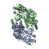
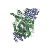
| Deposited unit | 
| ||||||||||||||||||||||||||||||
|---|---|---|---|---|---|---|---|---|---|---|---|---|---|---|---|---|---|---|---|---|---|---|---|---|---|---|---|---|---|---|---|
| 1 | 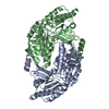
| ||||||||||||||||||||||||||||||
| 2 | 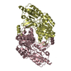
| ||||||||||||||||||||||||||||||
| Unit cell |
| ||||||||||||||||||||||||||||||
| Noncrystallographic symmetry (NCS) | NCS domain:
NCS domain segments:
|
-Components
| #1: Protein | / Ornithine-oxo-acid aminotransferase Mass: 47182.184 Da / Num. of mol.: 4 Source method: isolated from a genetically manipulated source Source: (gene. exp.) Plasmodium falciparum (malaria parasite P. falciparum)Strain: isolate CDC / Honduras / Gene: OAT / Plasmid: pET28a / Production host:  Escherichia coli (E. coli) / Strain (production host): BL21 / References: UniProt: Q07805, ornithine aminotransferase Escherichia coli (E. coli) / Strain (production host): BL21 / References: UniProt: Q07805, ornithine aminotransferase#2: Water | ChemComp-HOH / | Water Mass: 18.015 Da / Num. of mol.: 400 / Source method: isolated from a natural source / Formula: H2O Mass: 18.015 Da / Num. of mol.: 400 / Source method: isolated from a natural source / Formula: H2O |
|---|
-Experimental details
-Experiment
| Experiment | Method: X-RAY DIFFRACTION / Number of used crystals: 1 |
|---|
- Sample preparation
| Crystal | Density Matthews: 2.11 Å3/Da / Density % sol: 41.59 % |
|---|---|
| Crystal grow | Temperature: 293 K / Method: vapor diffusion, hanging drop / pH: 6 Details: 0.9 M lithium chloride, 0.1 M citric acid, 20 % PEG 6000, pH 6.0, VAPOR DIFFUSION, HANGING DROP, temperature 293K |
-Data collection
| Diffraction | Mean temperature: 100 K |
|---|---|
| Diffraction source | Source: SYNCHROTRON / Site: SLS  / Beamline: X06SA / Wavelength: 0.98696 Å / Beamline: X06SA / Wavelength: 0.98696 Å |
| Detector | Type: MAR CCD 165 mm / Detector: CCD / Date: Nov 28, 2008 / Details: mirrors |
| Radiation | Monochromator: mirrors / Protocol: SINGLE WAVELENGTH / Monochromatic (M) / Laue (L): M / Scattering type: x-ray |
| Radiation wavelength | Wavelength: 0.98696 Å / Relative weight: 1 |
| Reflection | Resolution: 2.3→43.4 Å / Num. all: 69479 / Num. obs: 66243 / % possible obs: 95.3 % / Observed criterion σ(F): 0 / Observed criterion σ(I): -3 / Redundancy: 2.2 % / Biso Wilson estimate: 45.4 Å2 / Rsym value: 0.05 / Net I/σ(I): 12.3 |
| Reflection shell | Resolution: 2.3→2.36 Å / Redundancy: 2.3 % / Mean I/σ(I) obs: 7.6 / Rsym value: 0.13 / % possible all: 10 |
- Processing
| Software |
| |||||||||||||||||||||||||||||||||||||||||||||||||||||||||||||||||||||||||||||||||||||||||||||||||||||||||||||||||||||||||||||||||||||||||||||||||||||||||||||||||||||||||||||||||||||||||||||||||||||||||||
|---|---|---|---|---|---|---|---|---|---|---|---|---|---|---|---|---|---|---|---|---|---|---|---|---|---|---|---|---|---|---|---|---|---|---|---|---|---|---|---|---|---|---|---|---|---|---|---|---|---|---|---|---|---|---|---|---|---|---|---|---|---|---|---|---|---|---|---|---|---|---|---|---|---|---|---|---|---|---|---|---|---|---|---|---|---|---|---|---|---|---|---|---|---|---|---|---|---|---|---|---|---|---|---|---|---|---|---|---|---|---|---|---|---|---|---|---|---|---|---|---|---|---|---|---|---|---|---|---|---|---|---|---|---|---|---|---|---|---|---|---|---|---|---|---|---|---|---|---|---|---|---|---|---|---|---|---|---|---|---|---|---|---|---|---|---|---|---|---|---|---|---|---|---|---|---|---|---|---|---|---|---|---|---|---|---|---|---|---|---|---|---|---|---|---|---|---|---|---|---|---|---|---|---|---|
| Refinement | Method to determine structure: MOLECULAR REPLACEMENT Starting model: pdb entry 1z7d Resolution: 2.3→43.375 Å / SU ML: 0.3 / Isotropic thermal model: overall / σ(F): 2 / Phase error: 26.92 / Stereochemistry target values: ML
| |||||||||||||||||||||||||||||||||||||||||||||||||||||||||||||||||||||||||||||||||||||||||||||||||||||||||||||||||||||||||||||||||||||||||||||||||||||||||||||||||||||||||||||||||||||||||||||||||||||||||||
| Solvent computation | Shrinkage radii: 0.9 Å / VDW probe radii: 1.11 Å / Solvent model: FLAT BULK SOLVENT MODEL / Bsol: 51.677 Å2 / ksol: 0.382 e/Å3 | |||||||||||||||||||||||||||||||||||||||||||||||||||||||||||||||||||||||||||||||||||||||||||||||||||||||||||||||||||||||||||||||||||||||||||||||||||||||||||||||||||||||||||||||||||||||||||||||||||||||||||
| Displacement parameters |
| |||||||||||||||||||||||||||||||||||||||||||||||||||||||||||||||||||||||||||||||||||||||||||||||||||||||||||||||||||||||||||||||||||||||||||||||||||||||||||||||||||||||||||||||||||||||||||||||||||||||||||
| Refine analyze | Luzzati coordinate error obs: 0.3 Å | |||||||||||||||||||||||||||||||||||||||||||||||||||||||||||||||||||||||||||||||||||||||||||||||||||||||||||||||||||||||||||||||||||||||||||||||||||||||||||||||||||||||||||||||||||||||||||||||||||||||||||
| Refinement step | Cycle: LAST / Resolution: 2.3→43.375 Å
| |||||||||||||||||||||||||||||||||||||||||||||||||||||||||||||||||||||||||||||||||||||||||||||||||||||||||||||||||||||||||||||||||||||||||||||||||||||||||||||||||||||||||||||||||||||||||||||||||||||||||||
| Refine LS restraints |
| |||||||||||||||||||||||||||||||||||||||||||||||||||||||||||||||||||||||||||||||||||||||||||||||||||||||||||||||||||||||||||||||||||||||||||||||||||||||||||||||||||||||||||||||||||||||||||||||||||||||||||
| Refine LS restraints NCS |
| |||||||||||||||||||||||||||||||||||||||||||||||||||||||||||||||||||||||||||||||||||||||||||||||||||||||||||||||||||||||||||||||||||||||||||||||||||||||||||||||||||||||||||||||||||||||||||||||||||||||||||
| LS refinement shell |
|
