[English] 日本語
Yorodumi
- PDB-2vqi: Structure of the P pilus usher (PapC) translocation pore -

+
Open data


ID or keywords:

Loading...

-
Basic information

Entry
Database: PDB / ID: 2vqi
TitleStructure of the P pilus usher (PapC) translocation pore
ComponentsOUTER MEMBRANE USHER PROTEIN PAPC
KeywordsTRANSPORT / TRANSMEMBRANE / OUTER MEMBRANE / USHER / P PILUS / MEMBRANE / FIMBRIUM / OM TRANSLOCATION PORE / PILUS ASSEMBLY PLATFORM
Function / homology
Function and homology information


fimbrial usher porin activity / pilus assembly / cell outer membrane / identical protein binding
Similarity search - Function
Outer membrane usher protein / Fimbrial membrane usher, conserved site / PapC, N-terminal domain / PapC, N-terminal domain superfamily / Outer membrane usher protein FimD, plug domain / PapC-like, C-terminal domain superfamily / Outer membrane usher protein / PapC C-terminal domain / PapC N-terminal domain / Fimbrial biogenesis outer membrane usher protein signature. / PapC-like, C-terminal domain
Similarity search - Domain/homology
Outer membrane usher protein PapC
Similarity search - Component
Biological speciesESCHERICHIA COLI (E. coli)
MethodX-RAY DIFFRACTION / SYNCHROTRON / MAD / Resolution: 3.2 Å
AuthorsRemaut, H. / Tang, C. / Henderson, N.S. / Pinkner, J.S. / Wang, T. / Hultgren, S.J. / Thanassi, D.G. / Li, H. / Waksman, G.
Citation
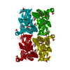
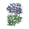
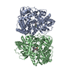
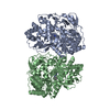
Journal: Cell / Year: 2008
Title: Fiber formation across the bacterial outer membrane by the chaperone/usher pathway.
Authors: Han Remaut / Chunyan Tang / Nadine S Henderson / Jerome S Pinkner / Tao Wang / Scott J Hultgren / David G Thanassi / Gabriel Waksman / Huilin Li /
Abstract: Gram-negative pathogens commonly exhibit adhesive pili on their surfaces that mediate specific attachment to the host. A major class of pili is assembled via the chaperone/usher pathway. Here, the ...Gram-negative pathogens commonly exhibit adhesive pili on their surfaces that mediate specific attachment to the host. A major class of pili is assembled via the chaperone/usher pathway. Here, the structural basis for pilus fiber assembly and secretion performed by the outer membrane assembly platform--the usher--is revealed by the crystal structure of the translocation domain of the P pilus usher PapC and single particle cryo-electron microscopy imaging of the FimD usher bound to a translocating type 1 pilus assembly intermediate. These structures provide molecular snapshots of a twinned-pore translocation machinery in action. Unexpectedly, only one pore is used for secretion, while both usher protomers are used for chaperone-subunit complex recruitment. The translocating pore itself comprises 24 beta strands and is occluded by a folded plug domain, likely gated by a conformationally constrained beta-hairpin. These structures capture the secretion of a virulence factor across the outer membrane of gram-negative bacteria.
History
DepositionMar 16, 2008Deposition site: PDBE / Processing site: PDBE
Revision 1.0May 27, 2008Provider: repository / Type: Initial release
Revision 1.1May 8, 2011Group: Version format compliance
Revision 1.2Jul 13, 2011Group: Version format compliance
Revision 1.3Jan 16, 2019Group: Data collection / Database references / Derived calculations
Category: citation / pdbx_struct_assembly ...citation / pdbx_struct_assembly / pdbx_struct_assembly_gen / pdbx_struct_assembly_prop
Item: _citation.country / _citation.journal_abbrev ..._citation.country / _citation.journal_abbrev / _citation.journal_id_ASTM / _citation.journal_id_CSD / _citation.journal_id_ISSN / _citation.page_last / _citation.pdbx_database_id_DOI / _citation.title
Remark 700 SHEET DETERMINATION METHOD: DSSP THE SHEETS PRESENTED AS "AA" IN EACH CHAIN ON SHEET RECORDS BELOW ... SHEET DETERMINATION METHOD: DSSP THE SHEETS PRESENTED AS "AA" IN EACH CHAIN ON SHEET RECORDS BELOW IS ACTUALLY AN 47-STRANDED BARREL THIS IS REPRESENTED BY A 48-STRANDED SHEET IN WHICH THE FIRST AND LAST STRANDS ARE IDENTICAL.

-
Structure visualization

Structure viewerMolecule:
MolmilJmol/JSmol

Downloads & links

-
Assembly

Deposited unit
A: OUTER MEMBRANE USHER PROTEIN PAPC
B: OUTER MEMBRANE USHER PROTEIN PAPC
hetero molecules


Theoretical massNumber of molelcules
Total (without water)116,3306
Polymers115,2582
Non-polymers1,0724
Water61334
1
A: OUTER MEMBRANE USHER PROTEIN PAPC
hetero molecules

A: OUTER MEMBRANE USHER PROTEIN PAPC
hetero molecules


Theoretical massNumber of molelcules
Total (without water)115,7174
Polymers115,2582
Non-polymers4592
Water362
TypeNameSymmetry operationNumber
identity operation1_555x,y,z1
crystal symmetry operation2_555-x,y,-z1
2
B: OUTER MEMBRANE USHER PROTEIN PAPC
hetero molecules

B: OUTER MEMBRANE USHER PROTEIN PAPC
hetero molecules


Theoretical massNumber of molelcules
Total (without water)116,9438
Polymers115,2582
Non-polymers1,6856
Water362
TypeNameSymmetry operationNumber
identity operation1_555x,y,z1
crystal symmetry operation2_555-x,y,-z1
Unit cell
Length a, b, c (Å)166.500, 101.900, 113.700
Angle α, β, γ (deg.)90.00, 128.20, 90.00
Int Tables number5
Space group name H-MC121
Noncrystallographic symmetry (NCS)NCS oper: (Code: given
Matrix: (0.2323, 0.0092, 0.9726), (0.0097, -0.9999, 0.0071), (0.9726, 0.0078, -0.2324)
Vector: 2.7337, 58.9495, -4.5976)

-
Components

#1: Protein OUTER MEMBRANE USHER PROTEIN PAPC / PAPC


Mass: 57628.984 Da / Num. of mol.: 2 / Fragment: TRANSLOCATION DOMAIN, RESIDUES 157-667
Source method: isolated from a genetically manipulated source
Source: (gene. exp.) ESCHERICHIA COLI (E. coli) / Strain: J96 / Production host: ESCHERICHIA COLI (E. coli) / Strain (production host): B834 (DE3) / References: UniProt: P07110
#2: Chemical ChemComp-LDA / LAURYL DIMETHYLAMINE-N-OXIDE / Lauryldimethylamine oxide


Mass: 229.402 Da / Num. of mol.: 2 / Source method: obtained synthetically / Formula: C14H31NO / Comment: LDAO, detergent*YM
#3: Chemical ChemComp-C8E / (HYDROXYETHYLOXY)TRI(ETHYLOXY)OCTANE


Mass: 306.438 Da / Num. of mol.: 2 / Source method: obtained synthetically / Formula: C16H34O5 / Comment: C8E, detergent*YM
#4: Water ChemComp-HOH / water / Water


Mass: 18.015 Da / Num. of mol.: 34 / Source method: isolated from a natural source / Formula: H2O
Sequence detailsCONSTRUCT CONTAINS CENTRAL TRANSLOCATION CHANNEL, RESIDUES 130-640. C-TERMINAL 4 RESIDUES LVPR ...CONSTRUCT CONTAINS CENTRAL TRANSLOCATION CHANNEL, RESIDUES 130-640. C-TERMINAL 4 RESIDUES LVPR RESULT FROM INTRODUCED THROMBIN CLEAVAGE SITE

-
Experimental details

-
Experiment

ExperimentMethod: X-RAY DIFFRACTION / Number of used crystals: 1

-
Sample preparation

CrystalDensity Matthews: 3.5 Å3/Da / Density % sol: 61.3 % / Description: NONE
Crystal growpH: 8.5
Details: 5-20 % 3-METHYL-1,5-PENTANEDIOL (MPD), 6-12 % POLYETHYLENE GLYCOL 4000 (PEG4000), 50 MM NA CITRATE PH 5.6, 100 MM AMMONIUM SULFATE

-
Data collection

DiffractionMean temperature: 100 K
Diffraction sourceSource: SYNCHROTRON / Site: ESRF / Beamline: ID29 / Wavelength: 0.979
DetectorType: ADSC CCD / Detector: CCD / Date: Jul 7, 2007
RadiationProtocol: SINGLE WAVELENGTH / Monochromatic (M) / Laue (L): M / Scattering type: x-ray
Radiation wavelengthWavelength: 0.979 Å / Relative weight: 1
ReflectionResolution: 3.2→20 Å / Num. obs: 48050 / % possible obs: 98.4 % / Observed criterion σ(I): -3 / Redundancy: 3.7 % / Biso Wilson estimate: 67.4 Å2 / Rmerge(I) obs: 0.1 / Net I/σ(I): 9.7
Reflection shellResolution: 3.2→3.4 Å / Redundancy: 3.8 % / Rmerge(I) obs: 0.22 / Mean I/σ(I) obs: 5.5 / % possible all: 99.3

-
Processing

Software
NameVersionClassification
CNS1.2refinement
XDSdata reduction
XSCALEdata scaling
SHARPphasing
RefinementMethod to determine structure: MAD
Starting model: NONE

Resolution: 3.2→15 Å / Rfactor Rfree error: 0.006 / Data cutoff high absF: 5055100.25 / Isotropic thermal model: RESTRAINED / Cross valid method: THROUGHOUT / σ(F): 0
Details: DISORDERED ATOMS IN PARTIALLY ORDERED RESIDUES WERE MODELED WITH OCCUPANCY 0.01
RfactorNum. reflection% reflectionSelection details
Rfree0.2958 2410 5 %RANDOM
Rwork0.2593 ---
obs0.2593 47937 99.9 %-
Solvent computationSolvent model: FLAT MODEL / Bsol: 20.4143 Å2 / ksol: 0.3 e/Å3
Displacement parametersBiso mean: 50.3 Å2
Baniso -1Baniso -2Baniso -3
1-7.369 Å20 Å219.591 Å2
2---6.277 Å20 Å2
3----1.092 Å2
Refine analyze
FreeObs
Luzzati coordinate error0.51 Å0.42 Å
Luzzati d res low-5 Å
Luzzati sigma a0.5 Å0.41 Å
Refinement stepCycle: LAST / Resolution: 3.2→15 Å
ProteinNucleic acidLigandSolventTotal
Num. atoms7517 0 74 34 7625
Refine LS restraints
Refine-IDTypeDev idealDev ideal target
X-RAY DIFFRACTIONc_bond_d0.016733
X-RAY DIFFRACTIONc_bond_d_na
X-RAY DIFFRACTIONc_bond_d_prot
X-RAY DIFFRACTIONc_angle_d
X-RAY DIFFRACTIONc_angle_d_na
X-RAY DIFFRACTIONc_angle_d_prot
X-RAY DIFFRACTIONc_angle_deg1.73555
X-RAY DIFFRACTIONc_angle_deg_na
X-RAY DIFFRACTIONc_angle_deg_prot
X-RAY DIFFRACTIONc_dihedral_angle_d29.1
X-RAY DIFFRACTIONc_dihedral_angle_d_na
X-RAY DIFFRACTIONc_dihedral_angle_d_prot
X-RAY DIFFRACTIONc_improper_angle_d1.57
X-RAY DIFFRACTIONc_improper_angle_d_na
X-RAY DIFFRACTIONc_improper_angle_d_prot
X-RAY DIFFRACTIONc_mcbond_it1.491.5
X-RAY DIFFRACTIONc_mcangle_it2.72
X-RAY DIFFRACTIONc_scbond_it2.592
X-RAY DIFFRACTIONc_scangle_it3.662.5
LS refinement shellResolution: 3.2→3.4 Å / Rfactor Rfree error: 0.017 / Total num. of bins used: 6
RfactorNum. reflection% reflection
Rfree0.323 384 4.8 %
Rwork0.291 7597 -
obs--100 %
Xplor file
Refine-IDSerial noParam fileTopol file
X-RAY DIFFRACTION1PROTEIN_REP.PARAMPROTEIN.TOP
X-RAY DIFFRACTION2WATER_REP.PARAMWATER.TOP
X-RAY DIFFRACTION3LDAO.PARAMLDAO.TOPOL
X-RAY DIFFRACTION4C8E4.PARAMC8E4.TOPOL

+
About Yorodumi

-
News

-
Feb 9, 2022. New format data for meta-information of EMDB entries

New format data for meta-information of EMDB entries

  • Version 3 of the EMDB header file is now the official format.
  • The previous official version 1.9 will be removed from the archive.

Related info.:EMDB header

External links:wwPDB to switch to version 3 of the EMDB data model

-
Aug 12, 2020. Covid-19 info

Covid-19 info

URL: https://pdbj.org/emnavi/covid19.php

New page: Covid-19 featured information page in EM Navigator.

Related info.:Covid-19 info / Mar 5, 2020. Novel coronavirus structure data

+
Mar 5, 2020. Novel coronavirus structure data

Novel coronavirus structure data

Related info.:Yorodumi Speices / Aug 12, 2020. Covid-19 info

External links:COVID-19 featured content - PDBj / Molecule of the Month (242):Coronavirus Proteases

+
Jan 31, 2019. EMDB accession codes are about to change! (news from PDBe EMDB page)

EMDB accession codes are about to change! (news from PDBe EMDB page)

  • The allocation of 4 digits for EMDB accession codes will soon come to an end. Whilst these codes will remain in use, new EMDB accession codes will include an additional digit and will expand incrementally as the available range of codes is exhausted. The current 4-digit format prefixed with “EMD-” (i.e. EMD-XXXX) will advance to a 5-digit format (i.e. EMD-XXXXX), and so on. It is currently estimated that the 4-digit codes will be depleted around Spring 2019, at which point the 5-digit format will come into force.
  • The EM Navigator/Yorodumi systems omit the EMD- prefix.

Related info.:Q: What is EMD? / ID/Accession-code notation in Yorodumi/EM Navigator

External links:EMDB Accession Codes are Changing Soon! / Contact to PDBj

+
Jul 12, 2017. Major update of PDB

Major update of PDB

  • wwPDB released updated PDB data conforming to the new PDBx/mmCIF dictionary.
  • This is a major update changing the version number from 4 to 5, and with Remediation, in which all the entries are updated.
  • In this update, many items about electron microscopy experimental information are reorganized (e.g. em_software).
  • Now, EM Navigator and Yorodumi are based on the updated data.

External links:wwPDB Remediation / Enriched Model Files Conforming to OneDep Data Standards Now Available in the PDB FTP Archive

-
Yorodumi

Thousand views of thousand structures

  • Yorodumi is a browser for structure data from EMDB, PDB, SASBDB, etc.
  • This page is also the successor to EM Navigator detail page, and also detail information page/front-end page for Omokage search.
  • The word "yorodu" (or yorozu) is an old Japanese word meaning "ten thousand". "mi" (miru) is to see.

Related info.:EMDB / PDB / SASBDB / Comparison of 3 databanks / Yorodumi Search / Aug 31, 2016. New EM Navigator & Yorodumi / Yorodumi Papers / Jmol/JSmol / Function and homology information / Changes in new EM Navigator and Yorodumi

Read more