[English] 日本語
Yorodumi
- PDB-5vm4: The apo form of the triclocarban-binding single domain camelid na... -

+
Open data


ID or keywords:

Loading...

-
Basic information

Entry
Database: PDB / ID: 5vm4
TitleThe apo form of the triclocarban-binding single domain camelid nanobody VHH T10
ComponentsSingle domain camelid nanobody VHH T10
KeywordsUNKNOWN FUNCTION / nanobody / triclocarban
Function / homologyACETATE ION / FORMIC ACID
Function and homology information
Biological speciesLama glama (llama)
MethodX-RAY DIFFRACTION / SYNCHROTRON / MOLECULAR REPLACEMENT / Resolution: 1.9 Å
AuthorsTabares-da Rosa, S. / Gonzalez-Sapienza, G. / Wilson, D.K.
CitationJournal: J. Mol. Recognit. / Year: 2019
Title: Structure and specificity of several triclocarban-binding single domain camelid antibody fragments.
Authors: Tabares-da Rosa, S. / Wogulis, L.A. / Wogulis, M.D. / Gonzalez-Sapienza, G. / Wilson, D.K.
History
DepositionApr 26, 2017Deposition site: RCSB / Processing site: RCSB
Revision 1.0May 2, 2018Provider: repository / Type: Initial release
Revision 1.1Nov 21, 2018Group: Data collection / Database references / Category: citation / citation_author
Item: _citation.country / _citation.journal_abbrev ..._citation.country / _citation.journal_abbrev / _citation.journal_id_CSD / _citation.journal_id_ISSN / _citation.page_first / _citation.page_last / _citation.pdbx_database_id_DOI / _citation.pdbx_database_id_PubMed / _citation.title / _citation.year
Revision 1.2Dec 26, 2018Group: Data collection / Database references / Category: citation / Item: _citation.journal_volume / _citation.year
Revision 1.3Apr 3, 2024Group: Data collection / Database references / Refinement description
Category: chem_comp_atom / chem_comp_bond ...chem_comp_atom / chem_comp_bond / database_2 / pdbx_initial_refinement_model
Item: _database_2.pdbx_DOI / _database_2.pdbx_database_accession

-
Structure visualization

Structure viewerMolecule:
MolmilJmol/JSmol

Downloads & links

-
Assembly

Deposited unit
A: Single domain camelid nanobody VHH T10
B: Single domain camelid nanobody VHH T10
C: Single domain camelid nanobody VHH T10
D: Single domain camelid nanobody VHH T10
E: Single domain camelid nanobody VHH T10
F: Single domain camelid nanobody VHH T10
G: Single domain camelid nanobody VHH T10
H: Single domain camelid nanobody VHH T10
I: Single domain camelid nanobody VHH T10
J: Single domain camelid nanobody VHH T10
K: Single domain camelid nanobody VHH T10
L: Single domain camelid nanobody VHH T10
hetero molecules


Theoretical massNumber of molelcules
Total (without water)181,75524
Polymers181,12512
Non-polymers63012
Water7,927440
1
A: Single domain camelid nanobody VHH T10
B: Single domain camelid nanobody VHH T10
hetero molecules


Theoretical massNumber of molelcules
Total (without water)30,3986
Polymers30,1872
Non-polymers2104
Water362
TypeNameSymmetry operationNumber
identity operation1_555x,y,z1
Buried area1640 Å2
ΔGint-14 kcal/mol
Surface area12960 Å2
MethodPISA
2
F: Single domain camelid nanobody VHH T10
hetero molecules

C: Single domain camelid nanobody VHH T10
hetero molecules


Theoretical massNumber of molelcules
Total (without water)30,2934
Polymers30,1872
Non-polymers1052
Water362
TypeNameSymmetry operationNumber
identity operation1_555x,y,z1
crystal symmetry operation4_546-x+1/2,y-1/2,-z+11
Buried area1610 Å2
ΔGint-13 kcal/mol
Surface area13140 Å2
MethodPISA
3
D: Single domain camelid nanobody VHH T10
I: Single domain camelid nanobody VHH T10
hetero molecules


Theoretical massNumber of molelcules
Total (without water)30,2473
Polymers30,1872
Non-polymers591
Water362
TypeNameSymmetry operationNumber
identity operation1_555x,y,z1
Buried area1600 Å2
ΔGint-12 kcal/mol
Surface area12990 Å2
MethodPISA
4
E: Single domain camelid nanobody VHH T10
J: Single domain camelid nanobody VHH T10
hetero molecules


Theoretical massNumber of molelcules
Total (without water)30,2473
Polymers30,1872
Non-polymers591
Water362
TypeNameSymmetry operationNumber
identity operation1_555x,y,z1
Buried area1580 Å2
ΔGint-13 kcal/mol
Surface area12840 Å2
MethodPISA
5
G: Single domain camelid nanobody VHH T10
hetero molecules

H: Single domain camelid nanobody VHH T10
hetero molecules


Theoretical massNumber of molelcules
Total (without water)30,2804
Polymers30,1872
Non-polymers922
Water362
TypeNameSymmetry operationNumber
identity operation1_555x,y,z1
crystal symmetry operation4_555-x+1/2,y+1/2,-z1
Buried area1620 Å2
ΔGint-14 kcal/mol
Surface area12950 Å2
MethodPISA
6
K: Single domain camelid nanobody VHH T10
L: Single domain camelid nanobody VHH T10
hetero molecules


Theoretical massNumber of molelcules
Total (without water)30,2934
Polymers30,1872
Non-polymers1052
Water362
TypeNameSymmetry operationNumber
identity operation1_555x,y,z1
Buried area1580 Å2
ΔGint-11 kcal/mol
Surface area12970 Å2
MethodPISA
Unit cell
Length a, b, c (Å)161.566, 93.254, 110.576
Angle α, β, γ (deg.)90.00, 104.07, 90.00
Int Tables number5
Space group name H-MC121

-
Components

#1: Antibody
Single domain camelid nanobody VHH T10


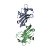
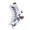
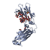
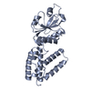
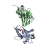
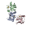
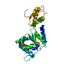
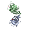
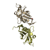
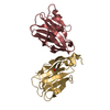
Mass: 15093.737 Da / Num. of mol.: 12
Source method: isolated from a genetically manipulated source
Source: (gene. exp.) Lama glama (llama) / Production host: Escherichia coli (E. coli)
#2: Chemical
ChemComp-ACT / ACETATE ION / Acetate


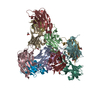
Mass: 59.044 Da / Num. of mol.: 6 / Source method: obtained synthetically / Formula: C2H3O2
#3: Chemical
ChemComp-FMT / FORMIC ACID / Formic acid


Mass: 46.025 Da / Num. of mol.: 6 / Source method: obtained synthetically / Formula: CH2O2
#4: Water ChemComp-HOH / water / Water


Mass: 18.015 Da / Num. of mol.: 440 / Source method: isolated from a natural source / Formula: H2O

-
Experimental details

-
Experiment

ExperimentMethod: X-RAY DIFFRACTION / Number of used crystals: 1

-
Sample preparation

CrystalDensity Matthews: 2.23 Å3/Da / Density % sol: 44.86 %
Crystal growTemperature: 298 K / Method: vapor diffusion, hanging drop / pH: 7.5
Details: protein concentration: 21 mg/ml well: 1.8305 M malonic acid, 0.25 M ammonium citrate tribasic, 0.12 M succinic acid, 0.3 M DL-malic acid, 0.4 M sodium acetate trihydrate, 0.5 M sodium ...Details: protein concentration: 21 mg/ml well: 1.8305 M malonic acid, 0.25 M ammonium citrate tribasic, 0.12 M succinic acid, 0.3 M DL-malic acid, 0.4 M sodium acetate trihydrate, 0.5 M sodium formate, 0.16 M ammonium tartrate dibasic

-
Data collection

DiffractionMean temperature: 100 K
Diffraction sourceSource: SYNCHROTRON / Site: SSRL / Beamline: BL9-2 / Wavelength: 0.97946 Å
DetectorType: DECTRIS PILATUS 6M / Detector: CCD / Date: Feb 24, 2017
RadiationProtocol: SINGLE WAVELENGTH / Monochromatic (M) / Laue (L): M / Scattering type: x-ray
Radiation wavelengthWavelength: 0.97946 Å / Relative weight: 1
ReflectionResolution: 1.9→107.26 Å / Num. obs: 120758 / % possible obs: 96.6 % / Redundancy: 2.9 % / Rmerge(I) obs: 0.077 / Net I/σ(I): 10
Reflection shellResolution: 1.9→1.93 Å / Rmerge(I) obs: 0.281 / Mean I/σ(I) obs: 4.3 / % possible all: 90.8

-
Processing

Software
NameVersionClassification
REFMAC5.8.0073refinement
XDSdata reduction
Aimlessdata scaling
PHASERphasing
RefinementMethod to determine structure: MOLECULAR REPLACEMENT
Starting model: T10 holo subunit A

Resolution: 1.9→40 Å / Cor.coef. Fo:Fc: 0.93 / Cor.coef. Fo:Fc free: 0.9 / SU B: 3.564 / SU ML: 0.106 / Cross valid method: THROUGHOUT / ESU R: 0.171 / ESU R Free: 0.156 / Details: HYDROGENS HAVE BEEN ADDED IN THE RIDING POSITIONS
RfactorNum. reflection% reflectionSelection details
Rfree0.2409 6051 5 %RANDOM
Rwork0.19696 ---
obs0.19914 114707 96.45 %-
Solvent computationIon probe radii: 0.8 Å / Shrinkage radii: 0.8 Å / VDW probe radii: 1.2 Å
Displacement parametersBiso mean: 15.835 Å2
Baniso -1Baniso -2Baniso -3
1--1.18 Å20 Å20.2 Å2
2---1.27 Å20 Å2
3---2.09 Å2
Refinement stepCycle: 1 / Resolution: 1.9→40 Å
ProteinNucleic acidLigandSolventTotal
Num. atoms12144 0 42 440 12626
Refine LS restraints
Refine-IDTypeDev idealDev ideal targetNumber
X-RAY DIFFRACTIONr_bond_refined_d0.0130.01912498
X-RAY DIFFRACTIONr_bond_other_d0.0010.0211256
X-RAY DIFFRACTIONr_angle_refined_deg1.5531.92116920
X-RAY DIFFRACTIONr_angle_other_deg0.769325872
X-RAY DIFFRACTIONr_dihedral_angle_1_deg6.18851560
X-RAY DIFFRACTIONr_dihedral_angle_2_deg31.14922.34564
X-RAY DIFFRACTIONr_dihedral_angle_3_deg14.944151944
X-RAY DIFFRACTIONr_dihedral_angle_4_deg20.06715108
X-RAY DIFFRACTIONr_chiral_restr0.0940.21752
X-RAY DIFFRACTIONr_gen_planes_refined0.0080.02114382
X-RAY DIFFRACTIONr_gen_planes_other0.0010.023126
X-RAY DIFFRACTIONr_nbd_refined
X-RAY DIFFRACTIONr_nbd_other
X-RAY DIFFRACTIONr_nbtor_refined
X-RAY DIFFRACTIONr_nbtor_other
X-RAY DIFFRACTIONr_xyhbond_nbd_refined
X-RAY DIFFRACTIONr_xyhbond_nbd_other
X-RAY DIFFRACTIONr_metal_ion_refined
X-RAY DIFFRACTIONr_metal_ion_other
X-RAY DIFFRACTIONr_symmetry_vdw_refined
X-RAY DIFFRACTIONr_symmetry_vdw_other
X-RAY DIFFRACTIONr_symmetry_hbond_refined
X-RAY DIFFRACTIONr_symmetry_hbond_other
X-RAY DIFFRACTIONr_symmetry_metal_ion_refined
X-RAY DIFFRACTIONr_symmetry_metal_ion_other
X-RAY DIFFRACTIONr_mcbond_it1.0611.4766282
X-RAY DIFFRACTIONr_mcbond_other1.0611.4766283
X-RAY DIFFRACTIONr_mcangle_it1.6432.2047824
X-RAY DIFFRACTIONr_mcangle_other1.6432.2047825
X-RAY DIFFRACTIONr_scbond_it1.6221.6566215
X-RAY DIFFRACTIONr_scbond_other1.6231.6566204
X-RAY DIFFRACTIONr_scangle_it
X-RAY DIFFRACTIONr_scangle_other2.5742.4039097
X-RAY DIFFRACTIONr_long_range_B_refined3.54911.87613706
X-RAY DIFFRACTIONr_long_range_B_other3.54911.87613707
X-RAY DIFFRACTIONr_rigid_bond_restr
X-RAY DIFFRACTIONr_sphericity_free
X-RAY DIFFRACTIONr_sphericity_bonded
LS refinement shellResolution: 1.9→1.95 Å / Total num. of bins used: 20
RfactorNum. reflection% reflection
Rfree0.281 433 -
Rwork0.236 7983 -
obs--91.83 %

+
About Yorodumi

-
News

-
Feb 9, 2022. New format data for meta-information of EMDB entries

New format data for meta-information of EMDB entries

  • Version 3 of the EMDB header file is now the official format.
  • The previous official version 1.9 will be removed from the archive.

Related info.:EMDB header

External links:wwPDB to switch to version 3 of the EMDB data model

-
Aug 12, 2020. Covid-19 info

Covid-19 info

URL: https://pdbj.org/emnavi/covid19.php

New page: Covid-19 featured information page in EM Navigator.

Related info.:Covid-19 info / Mar 5, 2020. Novel coronavirus structure data

+
Mar 5, 2020. Novel coronavirus structure data

Novel coronavirus structure data

Related info.:Yorodumi Speices / Aug 12, 2020. Covid-19 info

External links:COVID-19 featured content - PDBj / Molecule of the Month (242):Coronavirus Proteases

+
Jan 31, 2019. EMDB accession codes are about to change! (news from PDBe EMDB page)

EMDB accession codes are about to change! (news from PDBe EMDB page)

  • The allocation of 4 digits for EMDB accession codes will soon come to an end. Whilst these codes will remain in use, new EMDB accession codes will include an additional digit and will expand incrementally as the available range of codes is exhausted. The current 4-digit format prefixed with “EMD-” (i.e. EMD-XXXX) will advance to a 5-digit format (i.e. EMD-XXXXX), and so on. It is currently estimated that the 4-digit codes will be depleted around Spring 2019, at which point the 5-digit format will come into force.
  • The EM Navigator/Yorodumi systems omit the EMD- prefix.

Related info.:Q: What is EMD? / ID/Accession-code notation in Yorodumi/EM Navigator

External links:EMDB Accession Codes are Changing Soon! / Contact to PDBj

+
Jul 12, 2017. Major update of PDB

Major update of PDB

  • wwPDB released updated PDB data conforming to the new PDBx/mmCIF dictionary.
  • This is a major update changing the version number from 4 to 5, and with Remediation, in which all the entries are updated.
  • In this update, many items about electron microscopy experimental information are reorganized (e.g. em_software).
  • Now, EM Navigator and Yorodumi are based on the updated data.

External links:wwPDB Remediation / Enriched Model Files Conforming to OneDep Data Standards Now Available in the PDB FTP Archive

-
Yorodumi

Thousand views of thousand structures

  • Yorodumi is a browser for structure data from EMDB, PDB, SASBDB, etc.
  • This page is also the successor to EM Navigator detail page, and also detail information page/front-end page for Omokage search.
  • The word "yorodu" (or yorozu) is an old Japanese word meaning "ten thousand". "mi" (miru) is to see.

Related info.:EMDB / PDB / SASBDB / Comparison of 3 databanks / Yorodumi Search / Aug 31, 2016. New EM Navigator & Yorodumi / Yorodumi Papers / Jmol/JSmol / Function and homology information / Changes in new EM Navigator and Yorodumi

Read more