 Movie
Movie Controller

[English] 日本語
 Yorodumi
- PDB-5vlv: The apo form of the triclocarban-binding single domain camelid na... -
+ Open data

- Basic information
| Entry | Database: PDB / ID: 5vlv | ||||||
|---|---|---|---|---|---|---|---|
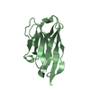
| Title | The apo form of the triclocarban-binding single domain camelid nanobody VHH T9 | ||||||
 Components Components | Single Domain Camelid Nanobody VHH T9 | ||||||
 Keywords Keywords | UNKNOWN FUNCTION /  nanobody / triclocarban nanobody / triclocarban | ||||||
| Biological species |  Lama glama (llama) Lama glama (llama) | ||||||
| Method | X-RAY DIFFRACTION / SYNCHROTRON / MOLECULAR REPLACEMENT / Resolution: 1.35 Å | ||||||
 Authors Authors | Tabares-da Rosa, S. / Gonzalez-Sapienza, G. / Wilson, D.K. | ||||||
 Citation Citation | Journal: J. Mol. Recognit. / Year: 2019 Title: Structure and specificity of several triclocarban-binding single domain camelid antibody fragments. Authors: Tabares-da Rosa, S. / Wogulis, L.A. / Wogulis, M.D. / Gonzalez-Sapienza, G. / Wilson, D.K. | ||||||
| History |
|
- Structure visualization
| Structure viewer | Molecule:  MolmilJmol/JSmol MolmilJmol/JSmol |
|---|
- Downloads & links
-Download
| PDBx/mmCIF format | 5vlv.cif.gz | 63.7 KB | Display | PDBx/mmCIF format |
|---|---|---|---|---|
| PDB format | pdb5vlv.ent.gz | 45.9 KB | Display | PDB format |
| PDBx/mmJSON format | 5vlv.json.gz | Tree view | PDBx/mmJSON format | |
| Others |  Other downloads Other downloads |
-Validation report
| Arichive directory | https://data.pdbj.org/pub/pdb/validation_reports/vl/5vlvftp://data.pdbj.org/pub/pdb/validation_reports/vl/5vlv | HTTPS FTP |
|---|
-Related structure data


-Links
 PDBj
PDBj


- Assembly
| Deposited unit | 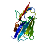
| ||||||||
|---|---|---|---|---|---|---|---|---|---|
| 1 |
| ||||||||
| Unit cell |
| ||||||||
| Components on special symmetry positions |
|
-Components
| #1: Antibody | Mass: 14289.056 Da / Num. of mol.: 1 Source method: isolated from a genetically manipulated source Source: (gene. exp.) Lama glama (llama) / Production host:  Escherichia coli (E. coli) Escherichia coli (E. coli) |
|---|---|
| #2: Water | ChemComp-HOH / Water Mass: 18.015 Da / Num. of mol.: 70 / Source method: isolated from a natural source / Formula: H2O Mass: 18.015 Da / Num. of mol.: 70 / Source method: isolated from a natural source / Formula: H2O |
-Experimental details
-Experiment
| Experiment | Method: X-RAY DIFFRACTION / Number of used crystals: 1 |
|---|
- Sample preparation
| Crystal | Density Matthews: 2.49 Å3/Da / Density % sol: 50.53 % |
|---|---|
| Crystal grow | Temperature: 298 K / Method: vapor diffusion, hanging drop / pH: 7.5 Details: protein concentration 40 mg/ml well 25% w/v PEG MME 3350, 200 mM ammonium sulfate, 100 mM Hepes |
-Data collection
| Diffraction | Mean temperature: 100 K |
|---|---|
| Diffraction source | Source: SYNCHROTRON / Site: ALS  / Beamline: 8.3.1 / Wavelength: 1.11583 Å / Beamline: 8.3.1 / Wavelength: 1.11583 Å |
| Detector | Type: DECTRIS PILATUS3 S 6M / Detector: CCD / Date: Jan 3, 2017 |
| Radiation | Protocol: SINGLE WAVELENGTH / Monochromatic (M) / Laue (L): M / Scattering type: x-ray |
| Radiation wavelength | Wavelength: 1.11583 Å / Relative weight: 1 |
| Reflection | Resolution: 1.35→48.81 Å / Num. obs: 32653 / % possible obs: 100 % / Redundancy: 6.8 % / Rmerge(I) obs: 0.031 / Net I/σ(I): 24.5 |
| Reflection shell | Resolution: 1.35→1.39 Å / Rmerge(I) obs: 0.624 / Mean I/σ(I) obs: 2.4 / % possible all: 99.9 |
- Processing
| Software |
| ||||||||||||||||||||||||||||||||||||||||||||||||||||||||||||||||||||||||||||||||||||||||||||||||||||||||||||||||||||||||||||||||||||||||||||||||||||||||||||||||||||||||||||||||||||||
|---|---|---|---|---|---|---|---|---|---|---|---|---|---|---|---|---|---|---|---|---|---|---|---|---|---|---|---|---|---|---|---|---|---|---|---|---|---|---|---|---|---|---|---|---|---|---|---|---|---|---|---|---|---|---|---|---|---|---|---|---|---|---|---|---|---|---|---|---|---|---|---|---|---|---|---|---|---|---|---|---|---|---|---|---|---|---|---|---|---|---|---|---|---|---|---|---|---|---|---|---|---|---|---|---|---|---|---|---|---|---|---|---|---|---|---|---|---|---|---|---|---|---|---|---|---|---|---|---|---|---|---|---|---|---|---|---|---|---|---|---|---|---|---|---|---|---|---|---|---|---|---|---|---|---|---|---|---|---|---|---|---|---|---|---|---|---|---|---|---|---|---|---|---|---|---|---|---|---|---|---|---|---|---|
| Refinement | Method to determine structure: MOLECULAR REPLACEMENT Starting model: T9 holo Resolution: 1.35→48.81 Å / Cor.coef. Fo:Fc: 0.97 / Cor.coef. Fo:Fc free: 0.961 / SU B: 1.472 / SU ML: 0.027 / Cross valid method: THROUGHOUT / ESU R: 0.048 / ESU R Free: 0.047 / Details: HYDROGENS HAVE BEEN ADDED IN THE RIDING POSITIONS
| ||||||||||||||||||||||||||||||||||||||||||||||||||||||||||||||||||||||||||||||||||||||||||||||||||||||||||||||||||||||||||||||||||||||||||||||||||||||||||||||||||||||||||||||||||||||
| Solvent computation | Ion probe radii: 0.8 Å / Shrinkage radii: 0.8 Å / VDW probe radii: 1.2 Å | ||||||||||||||||||||||||||||||||||||||||||||||||||||||||||||||||||||||||||||||||||||||||||||||||||||||||||||||||||||||||||||||||||||||||||||||||||||||||||||||||||||||||||||||||||||||
| Displacement parameters | Biso mean: 25.44 Å2
| ||||||||||||||||||||||||||||||||||||||||||||||||||||||||||||||||||||||||||||||||||||||||||||||||||||||||||||||||||||||||||||||||||||||||||||||||||||||||||||||||||||||||||||||||||||||
| Refinement step | Cycle: 1 / Resolution: 1.35→48.81 Å
| ||||||||||||||||||||||||||||||||||||||||||||||||||||||||||||||||||||||||||||||||||||||||||||||||||||||||||||||||||||||||||||||||||||||||||||||||||||||||||||||||||||||||||||||||||||||
| Refine LS restraints |
|