 Movie
Movie Controller

[English] 日本語
 Yorodumi
- PDB-5yxk: High resolution crystal structure of Human B7-2 IgV domain in P21... -
+ Open data

- Basic information
| Entry | Database: PDB / ID: 5yxk | ||||||
|---|---|---|---|---|---|---|---|
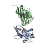
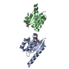
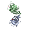
| Title | High resolution crystal structure of Human B7-2 IgV domain in P21 space group | ||||||
 Components Components | T-lymphocyte activation antigen CD86 | ||||||
 Keywords Keywords |  IMMUNE SYSTEM / B7-2 / co-stimulatory molecule / Ig super family / CD86 IMMUNE SYSTEM / B7-2 / co-stimulatory molecule / Ig super family / CD86 | ||||||
| Function / homology |  Function and homology information Function and homology informationpositive regulation of lymphotoxin A production / CD40 signaling pathway / positive regulation of T-helper 2 cell differentiation / activation of protein kinase C activity / CD28 co-stimulation / CD28 dependent Vav1 pathway / positive regulation of immunoglobulin production / CTLA4 inhibitory signaling / B cell activation / positive regulation of interleukin-4 production ...positive regulation of lymphotoxin A production / CD40 signaling pathway / positive regulation of T-helper 2 cell differentiation / activation of protein kinase C activity / CD28 co-stimulation / CD28 dependent Vav1 pathway / positive regulation of immunoglobulin production / CTLA4 inhibitory signaling / B cell activation / positive regulation of interleukin-4 production / Interleukin-10 signaling / CD28 dependent PI3K/Akt signaling / : / centriolar satellite / coreceptor activity / positive regulation of T cell proliferation / negative regulation of T cell proliferation / T cell costimulation / T cell activation / positive regulation of interleukin-2 production / positive regulation of non-canonical NF-kappaB signal transduction / Constitutive Signaling by Aberrant PI3K in Cancer / virus receptor activity / PIP3 activates AKT signaling / signaling receptor activity / PI5P, PP2A and IER3 Regulate PI3K/AKT Signaling / adaptive immune response / cellular response to lipopolysaccharide / receptor ligand activity / cell surface receptor signaling pathway / immune response / external side of plasma membrane / positive regulation of cell population proliferation / positive regulation of DNA-templated transcription / cell surface / extracellular exosome / plasma membraneSimilarity search - Function | ||||||
| Biological species |  Homo sapiens (human) Homo sapiens (human) | ||||||
| Method | X-RAY DIFFRACTION / SYNCHROTRON / MOLECULAR REPLACEMENT / molecular replacement / Resolution: 1.9 Å | ||||||
 Authors Authors | Lankipalli, S. / Ramagopal, U.A. | ||||||
 Citation Citation | Journal: To be published Title: Comparitive structural analysis of human B7-2: New insights on its possible dimeric state Authors: Lankipalli, S. / Ramagopal, U.A. | ||||||
| History |
|
- Structure visualization
| Structure viewer | Molecule: MolmilJmol/JSmol |
|---|
- Downloads & links
-Download
| PDBx/mmCIF format | 5yxk.cif.gz | 102.6 KB | Display | PDBx/mmCIF format |
|---|---|---|---|---|
| PDB format | pdb5yxk.ent.gz | 78.3 KB | Display | PDB format |
| PDBx/mmJSON format | 5yxk.json.gz | Tree view | PDBx/mmJSON format | |
| Others |  Other downloads Other downloads |
-Validation report
| Arichive directory | https://data.pdbj.org/pub/pdb/validation_reports/yx/5yxkftp://data.pdbj.org/pub/pdb/validation_reports/yx/5yxk | HTTPS FTP |
|---|
-Related structure data
| Related structure data |  1ncnS S: Starting model for refinement |
|---|---|
| Similar structure data |


-Links
 PDBj
PDBj


- Assembly
| Deposited unit | 
| ||||||||
|---|---|---|---|---|---|---|---|---|---|
| 1 | 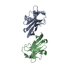
| ||||||||
| 2 | 
| ||||||||
| Unit cell |
| ||||||||
| Details | possible dimer |
-Components
| #1: Antibody | Mass: 12936.709 Da / Num. of mol.: 4 / Fragment: Ig-like V-type Source method: isolated from a genetically manipulated source Source: (gene. exp.) Homo sapiens (human) / Gene: CD86, CD28LG2 / Plasmid: pNIC28-Bsa4 / Production host:  Escherichia coli (E. coli) / Strain (production host): BL21(DE3) / References: UniProt: P42081 Escherichia coli (E. coli) / Strain (production host): BL21(DE3) / References: UniProt: P42081#2: Water | ChemComp-HOH / | Water Mass: 18.015 Da / Num. of mol.: 81 / Source method: isolated from a natural source / Formula: H2O Mass: 18.015 Da / Num. of mol.: 81 / Source method: isolated from a natural source / Formula: H2O |
|---|
-Experimental details
-Experiment
| Experiment | Method: X-RAY DIFFRACTION / Number of used crystals: 1 |
|---|
- Sample preparation
| Crystal | Density Matthews: 1.92 Å3/Da / Density % sol: 35.94 % / Mosaicity: 0.378 ° |
|---|---|
| Crystal grow | Temperature: 298 K / Method: vapor diffusion, sitting drop / pH: 8.5 Details: 0.1M Tris HCl pH 8.5, 2.4M Ammonium phosphate dibasic |
-Data collection
| Diffraction | Mean temperature: 100 K | |||||||||||||||||||||||||||||||||||||||||||||||||||||||||||||||||||||||||||||||||||||||||||||||||||||||||||||||||||||||||||||||||||||||||||||||||||||||||||||||||||||||||||||||||||||||||||||
|---|---|---|---|---|---|---|---|---|---|---|---|---|---|---|---|---|---|---|---|---|---|---|---|---|---|---|---|---|---|---|---|---|---|---|---|---|---|---|---|---|---|---|---|---|---|---|---|---|---|---|---|---|---|---|---|---|---|---|---|---|---|---|---|---|---|---|---|---|---|---|---|---|---|---|---|---|---|---|---|---|---|---|---|---|---|---|---|---|---|---|---|---|---|---|---|---|---|---|---|---|---|---|---|---|---|---|---|---|---|---|---|---|---|---|---|---|---|---|---|---|---|---|---|---|---|---|---|---|---|---|---|---|---|---|---|---|---|---|---|---|---|---|---|---|---|---|---|---|---|---|---|---|---|---|---|---|---|---|---|---|---|---|---|---|---|---|---|---|---|---|---|---|---|---|---|---|---|---|---|---|---|---|---|---|---|---|---|---|---|---|
| Diffraction source | Source: SYNCHROTRON / Site: ESRF  / Beamline: MASSIF-3 / Wavelength: 0.9677 Å / Beamline: MASSIF-3 / Wavelength: 0.9677 Å | |||||||||||||||||||||||||||||||||||||||||||||||||||||||||||||||||||||||||||||||||||||||||||||||||||||||||||||||||||||||||||||||||||||||||||||||||||||||||||||||||||||||||||||||||||||||||||||
| Detector | Type: DECTRIS EIGER X 4M / Detector: PIXEL / Date: Sep 29, 2017 | |||||||||||||||||||||||||||||||||||||||||||||||||||||||||||||||||||||||||||||||||||||||||||||||||||||||||||||||||||||||||||||||||||||||||||||||||||||||||||||||||||||||||||||||||||||||||||||
| Radiation | Protocol: SINGLE WAVELENGTH / Monochromatic (M) / Laue (L): M / Scattering type: x-ray | |||||||||||||||||||||||||||||||||||||||||||||||||||||||||||||||||||||||||||||||||||||||||||||||||||||||||||||||||||||||||||||||||||||||||||||||||||||||||||||||||||||||||||||||||||||||||||||
| Radiation wavelength | Wavelength: 0.9677 Å / Relative weight: 1 | |||||||||||||||||||||||||||||||||||||||||||||||||||||||||||||||||||||||||||||||||||||||||||||||||||||||||||||||||||||||||||||||||||||||||||||||||||||||||||||||||||||||||||||||||||||||||||||
| Reflection twin |
| |||||||||||||||||||||||||||||||||||||||||||||||||||||||||||||||||||||||||||||||||||||||||||||||||||||||||||||||||||||||||||||||||||||||||||||||||||||||||||||||||||||||||||||||||||||||||||||
| Reflection | Resolution: 1.9→50 Å / Num. obs: 30622 / % possible obs: 98.7 % / Redundancy: 4.1 % / Rmerge(I) obs: 0.107 / Rpim(I) all: 0.057 / Rrim(I) all: 0.122 / Χ2: 0.9 / Net I/σ(I): 7.3 | |||||||||||||||||||||||||||||||||||||||||||||||||||||||||||||||||||||||||||||||||||||||||||||||||||||||||||||||||||||||||||||||||||||||||||||||||||||||||||||||||||||||||||||||||||||||||||||
| Reflection shell | Diffraction-ID: 1
|
-Phasing
| Phasing | Method: molecular replacement |
|---|
- Processing
| Software |
| ||||||||||||||||||||||||||||||||||||||||||||||||||||||||||||
|---|---|---|---|---|---|---|---|---|---|---|---|---|---|---|---|---|---|---|---|---|---|---|---|---|---|---|---|---|---|---|---|---|---|---|---|---|---|---|---|---|---|---|---|---|---|---|---|---|---|---|---|---|---|---|---|---|---|---|---|---|---|
| Refinement | Method to determine structure: MOLECULAR REPLACEMENT Starting model: 1NCN Resolution: 1.9→36.74 Å / Cor.coef. Fo:Fc: 0.94 / Cor.coef. Fo:Fc free: 0.905 / SU B: 3.819 / SU ML: 0.117 / SU R Cruickshank DPI: 0.0426 / Cross valid method: THROUGHOUT / σ(F): 0 / ESU R: 0.043 / ESU R Free: 0.037 Details: HYDROGENS HAVE BEEN ADDED IN THE RIDING POSITIONS U VALUES : REFINED INDIVIDUALLY
| ||||||||||||||||||||||||||||||||||||||||||||||||||||||||||||
| Solvent computation | Ion probe radii: 0.8 Å / Shrinkage radii: 0.8 Å / VDW probe radii: 1.2 Å | ||||||||||||||||||||||||||||||||||||||||||||||||||||||||||||
| Displacement parameters | Biso max: 55.91 Å2 / Biso mean: 18.863 Å2 / Biso min: 7.77 Å2
| ||||||||||||||||||||||||||||||||||||||||||||||||||||||||||||
| Refinement step | Cycle: final / Resolution: 1.9→36.74 Å
| ||||||||||||||||||||||||||||||||||||||||||||||||||||||||||||
| Refine LS restraints |
| ||||||||||||||||||||||||||||||||||||||||||||||||||||||||||||
| LS refinement shell | Resolution: 1.885→1.934 Å / Rfactor Rfree error: 0 / Total num. of bins used: 20
|
