 Movie
Movie Controller

[English] 日本語
 Yorodumi
- PDB-4hag: Crystal structure of fc-fragment of human IgG2 antibody (centered... -
+ Open data

- Basic information
| Entry | Database: PDB / ID: 4hag | |||||||||
|---|---|---|---|---|---|---|---|---|---|---|
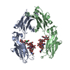
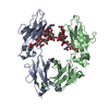
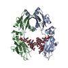
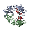
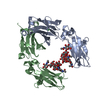
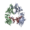
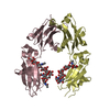
| Title | Crystal structure of fc-fragment of human IgG2 antibody (centered crystal form) | |||||||||
 Components Components | Ig gamma-2 chain C region | |||||||||
 Keywords Keywords |  IMMUNE SYSTEM / immunoglobulin fold IMMUNE SYSTEM / immunoglobulin fold | |||||||||
| Function / homology |  Function and homology information Function and homology informationClassical antibody-mediated complement activation / IgG immunoglobulin complex / Initial triggering of complement / FCGR activation / Role of phospholipids in phagocytosis / immunoglobulin complex, circulating / immunoglobulin receptor binding / complement activation, classical pathway / FCGR3A-mediated IL10 synthesis / antigen binding ...Classical antibody-mediated complement activation / IgG immunoglobulin complex / Initial triggering of complement / FCGR activation / Role of phospholipids in phagocytosis / immunoglobulin complex, circulating / immunoglobulin receptor binding / complement activation, classical pathway / FCGR3A-mediated IL10 synthesis / antigen binding / Regulation of Complement cascade / FCGR3A-mediated phagocytosis / B cell receptor signaling pathway / Regulation of actin dynamics for phagocytic cup formation / antibacterial humoral response / adaptive immune response / blood microparticle / extracellular space / extracellular exosome / extracellular region / plasma membraneSimilarity search - Function | |||||||||
| Biological species |  Homo sapiens (human) Homo sapiens (human) | |||||||||
| Method | X-RAY DIFFRACTION / MOLECULAR REPLACEMENT / Resolution: 3.4 Å | |||||||||
 Authors Authors | Teplyakov, A. / Malia, T. / Obmolova, G. / Zhao, Y. / Gilliland, G. | |||||||||
 Citation Citation | Journal: Mol.Immunol. / Year: 2013 Title: IgG2 Fc structure and the dynamic features of the IgG CH2-CH3 interface. Authors: Teplyakov, A. / Zhao, Y. / Malia, T.J. / Obmolova, G. / Gilliland, G.L. | |||||||||
| History |
|
- Structure visualization
| Structure viewer | Molecule: MolmilJmol/JSmol |
|---|
- Downloads & links
-Download
| PDBx/mmCIF format | 4hag.cif.gz | 109.4 KB | Display | PDBx/mmCIF format |
|---|---|---|---|---|
| PDB format | pdb4hag.ent.gz | 84.3 KB | Display | PDB format |
| PDBx/mmJSON format | 4hag.json.gz | Tree view | PDBx/mmJSON format | |
| Others |  Other downloads Other downloads |
-Validation report
| Arichive directory | https://data.pdbj.org/pub/pdb/validation_reports/ha/4hagftp://data.pdbj.org/pub/pdb/validation_reports/ha/4hag | HTTPS FTP |
|---|
-Related structure data
| Related structure data |  4hafC 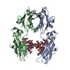 3aveS S: Starting model for refinement C: citing same article ( |
|---|---|
| Similar structure data |


-Links
 PDBj
PDBj


- Assembly
| Deposited unit | 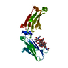
| ||||||||
|---|---|---|---|---|---|---|---|---|---|
| 1 | 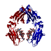
| ||||||||
| Unit cell |
|
-Components
| #1: Protein | Mass: 25112.469 Da / Num. of mol.: 1 / Fragment: CH2, CH3 DOMAINS Source method: isolated from a genetically manipulated source Source: (gene. exp.) Homo sapiens (human) / Gene: IGHG2 / Cell line (production host): HEK 293 / Production host: Homo sapiens (human) / References: UniProt: P01859 |
|---|---|
| #2: Polysaccharide | 2-acetamido-2-deoxy-beta-D-glucopyranose-(1-2)-alpha-D-mannopyranose-(1-3)-[2-acetamido-2-deoxy- ...2-acetamido-2-deoxy-beta-D-glucopyranose-(1-2)-alpha-D-mannopyranose-(1-3)-[2-acetamido-2-deoxy-beta-D-glucopyranose-(1-2)-alpha-D-mannopyranose-(1-6)]beta-D-mannopyranose-(1-4)-2-acetamido-2-deoxy-beta-D-glucopyranose-(1-4)-2-acetamido-2-deoxy-beta-D-glucopyranose / Mass: 1317.209 Da / Num. of mol.: 1 Source method: isolated from a genetically manipulated source |
| Sequence details | THERE IS A SER -> ALA SEQUENCE CONFLICT AT RESIDUE 257 IN UNIPROT DATABASE. |
-Experimental details
-Experiment
| Experiment | Method: X-RAY DIFFRACTION / Number of used crystals: 1 |
|---|
- Sample preparation
| Crystal | Density Matthews: 2.83 Å3/Da / Density % sol: 56.56 % |
|---|---|
| Crystal grow | Temperature: 293 K / Method: vapor diffusion, sitting drop / pH: 7 Details: 16% PEG 3350, 0.1 M HEPES. CRYO CONDITIONS: 23% PEG 3350, 0.1 M HEPES, 22% GLYCEROL, pH 7.0, VAPOR DIFFUSION, SITTING DROP, temperature 293K |
-Data collection
| Diffraction | Mean temperature: 100 K |
|---|---|
| Diffraction source | Source: ROTATING ANODE / Type: RIGAKU MICROMAX-007 HF / Wavelength: 1.5418 |
| Detector | Type: RIGAKU SATURN 944 / Detector: CCD / Date: Oct 6, 2009 / Details: VARIMAX HF |
| Radiation | Protocol: SINGLE WAVELENGTH / Monochromatic (M) / Laue (L): M / Scattering type: x-ray |
| Radiation wavelength | Wavelength: 1.5418 Å / Relative weight: 1 |
| Reflection | Resolution: 3.4→30 Å / Num. all: 3896 / Num. obs: 3896 / % possible obs: 94 % / Observed criterion σ(F): -3 / Observed criterion σ(I): -3 / Redundancy: 3.9 % / Biso Wilson estimate: 61.4 Å2 / Rmerge(I) obs: 0.084 / Net I/σ(I): 16.3 |
| Reflection shell | Resolution: 3.4→3.5 Å / Redundancy: 2.8 % / Rmerge(I) obs: 0.341 / Mean I/σ(I) obs: 3.1 / % possible all: 60.5 |
- Processing
| Software |
| |||||||||||||||||||||||||||||||||||||||||||||||||||||||||||||||||||||||||||
|---|---|---|---|---|---|---|---|---|---|---|---|---|---|---|---|---|---|---|---|---|---|---|---|---|---|---|---|---|---|---|---|---|---|---|---|---|---|---|---|---|---|---|---|---|---|---|---|---|---|---|---|---|---|---|---|---|---|---|---|---|---|---|---|---|---|---|---|---|---|---|---|---|---|---|---|---|
| Refinement | Method to determine structure: MOLECULAR REPLACEMENT Starting model: PDB entry 3ave Resolution: 3.4→15 Å / Cor.coef. Fo:Fc: 0.887 / Cor.coef. Fo:Fc free: 0.827 / SU B: 134.319 / SU ML: 0.854 / Cross valid method: THROUGHOUT / σ(F): 0 / ESU R Free: 0.836 / Stereochemistry target values: Engh & Huber
| |||||||||||||||||||||||||||||||||||||||||||||||||||||||||||||||||||||||||||
| Solvent computation | Ion probe radii: 0.8 Å / Shrinkage radii: 0.8 Å / VDW probe radii: 1.4 Å / Solvent model: BABINET MODEL WITH MASK | |||||||||||||||||||||||||||||||||||||||||||||||||||||||||||||||||||||||||||
| Displacement parameters | Biso mean: 193.5 Å2
| |||||||||||||||||||||||||||||||||||||||||||||||||||||||||||||||||||||||||||
| Refinement step | Cycle: LAST / Resolution: 3.4→15 Å
| |||||||||||||||||||||||||||||||||||||||||||||||||||||||||||||||||||||||||||
| Refine LS restraints |
| |||||||||||||||||||||||||||||||||||||||||||||||||||||||||||||||||||||||||||
| LS refinement shell | Resolution: 3.4→3.492 Å / Total num. of bins used: 20
| |||||||||||||||||||||||||||||||||||||||||||||||||||||||||||||||||||||||||||
| Refinement TLS params. | Method: refined / Refine-ID: X-RAY DIFFRACTION
| |||||||||||||||||||||||||||||||||||||||||||||||||||||||||||||||||||||||||||
| Refinement TLS group |
|
