 Movie
Movie Controller

[English] 日本語
 Yorodumi
- PDB-1fc1: CRYSTALLOGRAPHIC REFINEMENT AND ATOMIC MODELS OF A HUMAN FC FRAGM... -
+ Open data

- Basic information
| Entry | Database: PDB / ID: 1fc1 | |||||||||
|---|---|---|---|---|---|---|---|---|---|---|
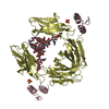
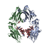
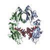
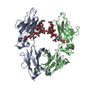
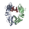
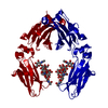
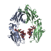
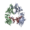
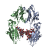
| Title | CRYSTALLOGRAPHIC REFINEMENT AND ATOMIC MODELS OF A HUMAN FC FRAGMENT AND ITS COMPLEX WITH FRAGMENT B OF PROTEIN A FROM STAPHYLOCOCCUS AUREUS AT 2.9-AND 2.8-ANGSTROMS RESOLUTION | |||||||||
 Components Components | FC FRAGMENT Fragment crystallizable region Fragment crystallizable region | |||||||||
 Keywords Keywords | IMMUNOGLOBULIN | |||||||||
| Function / homology |  Function and homology informationcomplement-dependent cytotoxicity / antibody-dependent cellular cytotoxicity / Fc-gamma receptor I complex binding / Classical antibody-mediated complement activation / Initial triggering of complement / immunoglobulin complex, circulating / IgG immunoglobulin complex / immunoglobulin receptor binding / FCGR activation / Role of phospholipids in phagocytosis ...complement-dependent cytotoxicity / antibody-dependent cellular cytotoxicity / Fc-gamma receptor I complex binding / Classical antibody-mediated complement activation / Initial triggering of complement / immunoglobulin complex, circulating / IgG immunoglobulin complex / immunoglobulin receptor binding / FCGR activation / Role of phospholipids in phagocytosis / complement activation, classical pathway / antigen binding / FCGR3A-mediated IL10 synthesis / Regulation of Complement cascade / FCGR3A-mediated phagocytosis / B cell receptor signaling pathway / Regulation of actin dynamics for phagocytic cup formation / antibacterial humoral response / Interleukin-4 and Interleukin-13 signaling / blood microparticle / adaptive immune response / extracellular space / extracellular exosome / extracellular region / plasma membrane Function and homology informationcomplement-dependent cytotoxicity / antibody-dependent cellular cytotoxicity / Fc-gamma receptor I complex binding / Classical antibody-mediated complement activation / Initial triggering of complement / immunoglobulin complex, circulating / IgG immunoglobulin complex / immunoglobulin receptor binding / FCGR activation / Role of phospholipids in phagocytosis ...complement-dependent cytotoxicity / antibody-dependent cellular cytotoxicity / Fc-gamma receptor I complex binding / Classical antibody-mediated complement activation / Initial triggering of complement / immunoglobulin complex, circulating / IgG immunoglobulin complex / immunoglobulin receptor binding / FCGR activation / Role of phospholipids in phagocytosis / complement activation, classical pathway / antigen binding / FCGR3A-mediated IL10 synthesis / Regulation of Complement cascade / FCGR3A-mediated phagocytosis / B cell receptor signaling pathway / Regulation of actin dynamics for phagocytic cup formation / antibacterial humoral response / Interleukin-4 and Interleukin-13 signaling / blood microparticle / adaptive immune response / extracellular space / extracellular exosome / extracellular region / plasma membraneSimilarity search - Function | |||||||||
| Biological species |  Homo sapiens (human) Homo sapiens (human) | |||||||||
| Method | X-RAY DIFFRACTION / Resolution: 2.9 Å | |||||||||
 Authors Authors | Deisenhofer, J. | |||||||||
 Citation Citation | Journal: Biochemistry / Year: 1981 Title: Crystallographic refinement and atomic models of a human Fc fragment and its complex with fragment B of protein A from Staphylococcus aureus at 2.9- and 2.8-A resolution. Authors: Deisenhofer, J. #1: Journal: Hoppe-Seyler's Z.Physiol.Chem. / Year: 1976Title: Crystallographic Structural Studies of a Human Fc Fragment. II. A Complete Model Based on a Fourier Map at 3.5 Angstroms Resolution Authors: Deisenhofer, J. / Colman, P.M. / Epp, O. / Huber, R. #2: Journal: Hoppe-Seyler's Z.Physiol.Chem. / Year: 1976Title: Crystallographic Structural Studies of a Human Fc-Fragment. I. An Electron-Density Map at 4 Angstroms Resolution and a Partial Model Authors: Deisenhofer, J. / Colman, P.M. / Huber, R. / Haupt, H. / Schwick, G. #3: Journal: FEBS Lett. / Year: 1974Title: X-Ray Studies on Antibody Fragments Authors: Colman, P.M. / Epp, O. / Fehlhammer, H. / Bode, W. / Schiffer, M. / Lattman, E.E. / Jones, T.A. | |||||||||
| History |
|
- Structure visualization
| Structure viewer | Molecule: MolmilJmol/JSmol |
|---|
- Downloads & links
-Download
| PDBx/mmCIF format | 1fc1.cif.gz | 87.2 KB | Display | PDBx/mmCIF format |
|---|---|---|---|---|
| PDB format | pdb1fc1.ent.gz | 72.9 KB | Display | PDB format |
| PDBx/mmJSON format | 1fc1.json.gz | Tree view | PDBx/mmJSON format | |
| Others |  Other downloads Other downloads |
-Validation report
| Arichive directory | https://data.pdbj.org/pub/pdb/validation_reports/fc/1fc1ftp://data.pdbj.org/pub/pdb/validation_reports/fc/1fc1 | HTTPS FTP |
|---|
-Related structure data


-Links
 PDBj
PDBj


- Assembly
| Deposited unit | 
| ||||||||
|---|---|---|---|---|---|---|---|---|---|
| 1 |
| ||||||||
| Unit cell |
| ||||||||
| Atom site foot note | 1: THESE ATOMS WERE NOT FOUND IN THE ELECTRON DENSITY MAP. THEIR COORDINATES WERE GENERATED USING STEREOCHEMICAL CRITERIA 2: RESIDUE 374 OF EACH CHAIN IS A CIS-PROLINE. | ||||||||
| Details | THE CRYSTALLOGRAPHIC ASYMMETRIC UNIT CONTAINS ONE FC FRAGMENT WHICH CONSISTS OF TWO CHEMICALLY IDENTICAL POLYPEPTIDE CHAINS EACH WITH ATTACHED POLYSACCHARIDE. THESE TWO CHAINS ARE DESIGNATED CHAIN 1 AND CHAIN 2 BY THE DEPOSITOR AND REPRESENTED WITH CHAIN IDENTIFIERS A AND B BELOW. THE CH2 AND CH3 DOMAINS OF THE TWO CRYSTALLOGRAPHICALLY INDEPENDENT POLYPEPTIDE CHAINS ARE RELATED BY NON-CRYSTALLOGRAPHIC (APPROXIMATE) DIADS (SEE JRNL REFERENCE ABOVE FOR A COMPLETE DISCUSSION). THE FOLLOWING TRANSFORMATION, WHEN APPLIED TO THE CH2 DOMAIN OF CHAIN A (RESIDUES PRO A 238 THROUGH GLY A 341 TOGETHER WITH THE POLYSACCHARIDE), WILL YIELD APPROXIMATE COORDINATES FOR THE CH2 DOMAIN OF CHAIN B (RESIDUES PRO B 238 THROUGH GLY B 341 TOGETHER WITH THE POLYSACCHARIDE). TRNSF1 1 -0.99391 -0.03853 -0.10322 92.070 TRNSF2 1 -0.06679 0.95579 0.28635 -0.310 TRNSF3 1 0.08759 0.29136 -0.95207 11.160 THE FOLLOWING TRANSFORMATION, WHEN APPLIED TO THE CH3 DOMAIN OF CHAIN A (RESIDUES GLN A 342 THROUGH LEU A 443), WILL YIELD APPROXIMATE COORDINATES FOR THE CH3 DOMAIN OF CHAIN B (RESIDUES GLN B 342 THROUGH LEU B 443). TRNSF1 2 -0.99907 -0.03950 -0.01721 88.510 TRNSF2 2 -0.04232 0.97468 0.21956 -1.010 TRNSF3 2 0.00810 0.22008 -0.97544 24.570 |
-Components
| #1: Protein | Fragment crystallizable region Mass: 25236.615 Da / Num. of mol.: 2 Source method: isolated from a genetically manipulated source Source: (gene. exp.) Homo sapiens (human) / Tissue: SERUM / References: UniProt: P01857#2: Polysaccharide | / Mass: 1625.490 Da / Num. of mol.: 2Source method: isolated from a genetically manipulated source |
|---|
-Experimental details
-Experiment
| Experiment | Method: X-RAY DIFFRACTION |
|---|
- Sample preparation
| Crystal | Density Matthews: 2.94 Å3/Da / Density % sol: 58.1 % |
|---|
-Data collection
| Radiation | Scattering type: x-ray |
|---|---|
| Radiation wavelength | Relative weight: 1 |
- Processing
| Refinement | Highest resolution: 2.9 Å | ||||||||||||
|---|---|---|---|---|---|---|---|---|---|---|---|---|---|
| Refinement step | Cycle: LAST / Highest resolution: 2.9 Å
|
