 Movie
Movie Controller


+ Open data

- Basic information
| Entry | Database: PDB / ID: 5tvo | |||||||||
|---|---|---|---|---|---|---|---|---|---|---|
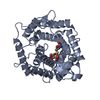
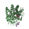
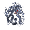
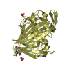
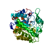
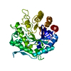
| Title | Crystal structure of Trypanosoma brucei AdoMetDC-delta26 monomer | |||||||||
 Components Components | (S-adenosylmethionine decarboxylase proenzyme) x 2 | |||||||||
 Keywords Keywords |  LYASE / autoinhibition / cis-proline LYASE / autoinhibition / cis-proline | |||||||||
| Function / homology |  Function and homology information Function and homology informationspermine biosynthetic process / adenosylmethionine decarboxylase / adenosylmethionine decarboxylase activity / spermidine biosynthetic process / cytosolSimilarity search - Function | |||||||||
| Biological species |  Trypanosoma brucei brucei (eukaryote) Trypanosoma brucei brucei (eukaryote) | |||||||||
| Method | X-RAY DIFFRACTION / SYNCHROTRON / MOLECULAR REPLACEMENT / Resolution: 1.481 Å | |||||||||
 Authors Authors | Volkov, O.A. / Ariagno, C. / Chen, Z. / Tomchick, D.R. / Phillips, M.A. | |||||||||
| Funding support |  United States, 2items United States, 2items
| |||||||||
 Citation Citation | Journal: Elife / Year: 2016 Title: Relief of autoinhibition by conformational switch explains enzyme activation by a catalytically dead paralog. Authors: Volkov, O.A. / Kinch, L.N. / Ariagno, C. / Deng, X. / Zhong, S. / Grishin, N.V. / Tomchick, D.R. / Chen, Z. / Phillips, M.A. | |||||||||
| History |
|
- Structure visualization
| Structure viewer | Molecule: MolmilJmol/JSmol |
|---|
- Downloads & links
-Download
| PDBx/mmCIF format | 5tvo.cif.gz | 201.1 KB | Display | PDBx/mmCIF format |
|---|---|---|---|---|
| PDB format | pdb5tvo.ent.gz | 161.7 KB | Display | PDB format |
| PDBx/mmJSON format | 5tvo.json.gz | Tree view | PDBx/mmJSON format | |
| Others |  Other downloads Other downloads |
-Validation report
| Arichive directory | https://data.pdbj.org/pub/pdb/validation_reports/tv/5tvoftp://data.pdbj.org/pub/pdb/validation_reports/tv/5tvo | HTTPS FTP |
|---|
-Related structure data
| Related structure data | 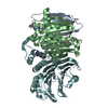 5tvfC  5tvmC 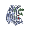 3ep9S C: citing same article ( S: Starting model for refinement |
|---|---|
| Similar structure data |


-Links
 PDBj
PDBj

- Assembly
| Deposited unit | 
| ||||||||
|---|---|---|---|---|---|---|---|---|---|
| 1 |
| ||||||||
| Unit cell |
|
-Components
| #1: Protein | Mass: 31971.945 Da / Num. of mol.: 1 Source method: isolated from a genetically manipulated source Source: (gene. exp.) Trypanosoma brucei brucei (strain 927/4 GUTat10.1) (eukaryote)Strain: 927/4 GUTat10.1 / Gene: Tb927.6.4410, Tb927.6.4460 / Plasmid: pET28bSmt3 / Production host:  Escherichia coli (E. coli) / Strain (production host): BL21 Escherichia coli (E. coli) / Strain (production host): BL21References: UniProt: Q587A7, adenosylmethionine decarboxylase |
|---|---|
| #2: Protein | Mass: 6782.649 Da / Num. of mol.: 1 / Fragment: UNP residues 27-85 Source method: isolated from a genetically manipulated source Source: (gene. exp.) Trypanosoma brucei brucei (strain 927/4 GUTat10.1) (eukaryote)Strain: 927/4 GUTat10.1 / Gene: Tb927.6.4410, Tb927.6.4460 / Production host: Escherichia coli (E. coli) / Strain (production host): BL21References: UniProt: Q587A7, adenosylmethionine decarboxylase |
| #3: Chemical | ChemComp-PYR / Pyruvic acid  Mass: 88.062 Da / Num. of mol.: 1 Mass: 88.062 Da / Num. of mol.: 1Source method: isolated from a genetically manipulated source Formula: C3H4O3 |
| #4: Chemical | ChemComp-NA /   Mass: 22.990 Da / Num. of mol.: 1 / Source method: obtained synthetically / Formula: Na Mass: 22.990 Da / Num. of mol.: 1 / Source method: obtained synthetically / Formula: Na |
| #5: Water | ChemComp-HOH / Water Mass: 18.015 Da / Num. of mol.: 252 / Source method: isolated from a natural source / Formula: H2O Mass: 18.015 Da / Num. of mol.: 252 / Source method: isolated from a natural source / Formula: H2O |
-Experimental details
-Experiment
| Experiment | Method: X-RAY DIFFRACTION / Number of used crystals: 1 |
|---|
- Sample preparation
| Crystal | Density Matthews: 2 Å3/Da / Density % sol: 38.45 % / Description: block |
|---|---|
| Crystal grow | Temperature: 293 K / Method: vapor diffusion, sitting drop / pH: 5.5 Details: 25 % PEG 3350, 0.2 M Ammonium acetate, 0.1 M Bis-tris, pH 5.5 |
-Data collection
| Diffraction | Mean temperature: 100 K |
|---|---|
| Diffraction source | Source: SYNCHROTRON / Site: APS / Beamline: 19-ID / Wavelength: 0.97935 Å |
| Detector | Type: ADSC QUANTUM 315 / Detector: CCD / Date: Jun 18, 2015 |
| Radiation | Monochromator: Graphite / Protocol: SINGLE WAVELENGTH / Monochromatic (M) / Laue (L): M / Scattering type: x-ray |
| Radiation wavelength | Wavelength: 0.97935 Å / Relative weight: 1 |
| Reflection | Resolution: 1.48→50 Å / Num. obs: 51575 / % possible obs: 98.2 % / Redundancy: 9.4 % / Biso Wilson estimate: 17.2 Å2 / Rsym value: 0.048 / Net I/σ(I): 29.8 |
| Reflection shell | Resolution: 1.48→1.51 Å / Redundancy: 4.8 % / Mean I/σ(I) obs: 1.4 / Num. unique all: 4315 / CC1/2: 0.68 / % possible all: 79.2 |
- Processing
| Software |
| |||||||||||||||||||||||||||||||||||||||||||||||||||||||||||||||||||||||||||||||||||||||||||||||||||||||||
|---|---|---|---|---|---|---|---|---|---|---|---|---|---|---|---|---|---|---|---|---|---|---|---|---|---|---|---|---|---|---|---|---|---|---|---|---|---|---|---|---|---|---|---|---|---|---|---|---|---|---|---|---|---|---|---|---|---|---|---|---|---|---|---|---|---|---|---|---|---|---|---|---|---|---|---|---|---|---|---|---|---|---|---|---|---|---|---|---|---|---|---|---|---|---|---|---|---|---|---|---|---|---|---|---|---|---|
| Refinement | Method to determine structure: MOLECULAR REPLACEMENT Starting model: 3EP9 Resolution: 1.481→38.006 Å / SU ML: 0.18 / Cross valid method: FREE R-VALUE / σ(F): 1.34 / Phase error: 22.28 / Stereochemistry target values: ML
| |||||||||||||||||||||||||||||||||||||||||||||||||||||||||||||||||||||||||||||||||||||||||||||||||||||||||
| Solvent computation | Shrinkage radii: 0.9 Å / VDW probe radii: 1.11 Å / Solvent model: FLAT BULK SOLVENT MODEL | |||||||||||||||||||||||||||||||||||||||||||||||||||||||||||||||||||||||||||||||||||||||||||||||||||||||||
| Refinement step | Cycle: LAST / Resolution: 1.481→38.006 Å
| |||||||||||||||||||||||||||||||||||||||||||||||||||||||||||||||||||||||||||||||||||||||||||||||||||||||||
| Refine LS restraints |
| |||||||||||||||||||||||||||||||||||||||||||||||||||||||||||||||||||||||||||||||||||||||||||||||||||||||||
| LS refinement shell |
|
