 Movie
Movie Controller

[English] 日本語
 Yorodumi
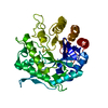
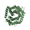
- PDB-6zbx: Structure of the catalytic domain of the Bacillus circulans alpha... -
+ Open data

- Basic information
| Entry | Database: PDB / ID: 6zbx | |||||||||
|---|---|---|---|---|---|---|---|---|---|---|
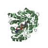
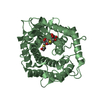
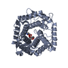
| Title | Structure of the catalytic domain of the Bacillus circulans alpha-1,6 Mannanase in complex with an alpha-1,6- alpha-manno-cyclophellitol carbasugar-stabilised trisaccharide inhibitor | |||||||||
 Components Components |
| |||||||||
 Keywords Keywords |  HYDROLASE / mannanase / cyclophellitol / epoxide HYDROLASE / mannanase / cyclophellitol / epoxide | |||||||||
| Function / homology |  Function and homology informationcatalytic activity / carbohydrate binding / carbohydrate metabolic process Function and homology informationcatalytic activity / carbohydrate binding / carbohydrate metabolic processSimilarity search - Function | |||||||||
| Biological species |  Bacillus circulans (bacteria) Bacillus circulans (bacteria) | |||||||||
| Method | X-RAY DIFFRACTION / SYNCHROTRON / MOLECULAR REPLACEMENT / Resolution: 1.35 Å | |||||||||
 Authors Authors | Schroeder, S. / Offen, W.A. / Jin, Y. / De Boer, C. / Enoterpi, J. / Marino, L. / van der Marel, G.A. / Codee, J.D.C. / Overkleeft, H.S. / Davies, G.J. | |||||||||
| Funding support |  United Kingdom, 2items United Kingdom, 2items
| |||||||||
 Citation Citation | Journal: Chemistry / Year: 2021 Title: Development of Non-Hydrolysable Oligosaccharide Activity-Based Inactivators for Endoglycanases: A Case Study on alpha-1,6 Mannanases. Authors: Schroder, S.P. / Offen, W.A. / Males, A. / Jin, Y. / de Boer, C. / Enotarpi, J. / Marino, L. / van der Marel, G.A. / Florea, B.I. / Codee, J.D.C. / Overkleeft, H.S. / Davies, G.J. | |||||||||
| History |
|
- Structure visualization
| Structure viewer | Molecule: MolmilJmol/JSmol |
|---|
- Downloads & links
-Download
| PDBx/mmCIF format | 6zbx.cif.gz | 309.5 KB | Display | PDBx/mmCIF format |
|---|---|---|---|---|
| PDB format | pdb6zbx.ent.gz | 246.8 KB | Display | PDB format |
| PDBx/mmJSON format | 6zbx.json.gz | Tree view | PDBx/mmJSON format | |
| Others |  Other downloads Other downloads |
-Validation report
| Arichive directory | https://data.pdbj.org/pub/pdb/validation_reports/zb/6zbxftp://data.pdbj.org/pub/pdb/validation_reports/zb/6zbx | HTTPS FTP |
|---|
-Related structure data
| Related structure data |  6zbmC  6zbwC  7nl5C  4d4aS S: Starting model for refinement C: citing same article ( |
|---|---|
| Similar structure data |


-Links
 PDBj
PDBj


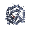
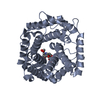
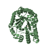
- Assembly
| Deposited unit | 
| ||||||||
|---|---|---|---|---|---|---|---|---|---|
| 1 | 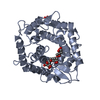
| ||||||||
| 2 | 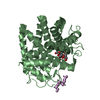
| ||||||||
| Unit cell |
|
-Components
-Protein / Protein/peptide / Sugars , 3 types, 5 molecules ABE
| #1: Protein | Mass: 40930.859 Da / Num. of mol.: 2 / Mutation: R341Q Source method: isolated from a genetically manipulated source Details: N-terminally His-tagged catalytic domain of BcGH76 with mutation R341Q Source: (gene. exp.) Bacillus circulans (bacteria) / Gene: aman6 / Production host: Escherichia coli (E. coli) / Variant (production host): Tuner (DE3) / References: UniProt: Q9Z4P9#2: Protein/peptide | | Mass: 358.434 Da / Num. of mol.: 1 Source method: isolated from a genetically manipulated source Details: Peptide, probably connected to either chain A or B, but unable to be identified Source: (gene. exp.) Bacillus circulans (bacteria) / Production host: Escherichia coli (E. coli)#6: Sugar | Mannose Type: D-saccharide, alpha linking / Mass: 180.156 Da / Num. of mol.: 2 Type: D-saccharide, alpha linking / Mass: 180.156 Da / Num. of mol.: 2Source method: isolated from a genetically manipulated source Formula: C6H12O6 |
|---|
-Non-polymers , 4 types, 589 molecules 


| #3: Chemical | Ethylene glycol Mass: 62.068 Da / Num. of mol.: 2 / Source method: obtained synthetically / Formula: C2H6O2 Mass: 62.068 Da / Num. of mol.: 2 / Source method: obtained synthetically / Formula: C2H6O2#4: Chemical |  Mass: 194.182 Da / Num. of mol.: 2 / Source method: obtained synthetically / Formula: C7H14O6 Mass: 194.182 Da / Num. of mol.: 2 / Source method: obtained synthetically / Formula: C7H14O6#5: Chemical |  Mass: 178.183 Da / Num. of mol.: 2 / Source method: obtained synthetically / Formula: C7H14O5 Mass: 178.183 Da / Num. of mol.: 2 / Source method: obtained synthetically / Formula: C7H14O5#7: Water | ChemComp-HOH / | WaterMass: 18.015 Da / Num. of mol.: 583 / Source method: isolated from a natural source / Formula: H2O |
|---|
-Details
| Has ligand of interest | Y |
|---|
-Experimental details
-Experiment
| Experiment | Method: X-RAY DIFFRACTION / Number of used crystals: 1 |
|---|
- Sample preparation
| Crystal | Density Matthews: 1.92 Å3/Da / Density % sol: 36.01 % |
|---|---|
| Crystal grow | Temperature: 291 K / Method: vapor diffusion, sitting drop / Details: PEG 3350, ammonium nitrate |
-Data collection
| Diffraction | Mean temperature: 100 K / Serial crystal experiment: N |
|---|---|
| Diffraction source | Source: SYNCHROTRON / Site: Diamond / Beamline: I04 / Wavelength: 0.9795 Å |
| Detector | Type: DECTRIS EIGER X 16M / Detector: PIXEL / Date: Feb 20, 2020 |
| Radiation | Protocol: SINGLE WAVELENGTH / Monochromatic (M) / Laue (L): M / Scattering type: x-ray |
| Radiation wavelength | Wavelength: 0.9795 Å / Relative weight: 1 |
| Reflection | Resolution: 1.35→50.49 Å / Num. obs: 127986 / % possible obs: 95.1 % / Redundancy: 3.5 % / CC1/2: 0.977 / Rpim(I) all: 0.026 / Net I/σ(I): 14.1 |
| Reflection shell | Resolution: 1.35→1.37 Å / Redundancy: 3 % / Num. unique obs: 5907 / CC1/2: 0.638 / Rpim(I) all: 0.398 / % possible all: 88.5 |
- Processing
| Software |
| |||||||||||||||||||||||||||||||||||||||||||||||||||||||||||||||||
|---|---|---|---|---|---|---|---|---|---|---|---|---|---|---|---|---|---|---|---|---|---|---|---|---|---|---|---|---|---|---|---|---|---|---|---|---|---|---|---|---|---|---|---|---|---|---|---|---|---|---|---|---|---|---|---|---|---|---|---|---|---|---|---|---|---|---|
| Refinement | Method to determine structure: MOLECULAR REPLACEMENT Starting model: 4D4A.PDB Resolution: 1.35→50.49 Å / Cor.coef. Fo:Fc: 0.979 / Cor.coef. Fo:Fc free: 0.965 / SU B: 2.458 / SU ML: 0.043 / Cross valid method: THROUGHOUT / σ(F): 0 / ESU R: 0.056 / ESU R Free: 0.056 / Stereochemistry target values: MAXIMUM LIKELIHOOD Details: THERE ARE REGIONS OF UNMODELLED DENSITY NEAR THE SIDE CHAINS OF PHE122A, ASN206A AND 206B, ASP266A AND TYR319A. THERE IS A PEPTIDE CHAIN OF 4 AMINO ACIDS WHICH COULD NOT BE ASSIGNED WITH ...Details: THERE ARE REGIONS OF UNMODELLED DENSITY NEAR THE SIDE CHAINS OF PHE122A, ASN206A AND 206B, ASP266A AND TYR319A. THERE IS A PEPTIDE CHAIN OF 4 AMINO ACIDS WHICH COULD NOT BE ASSIGNED WITH CONFIDENCE TO EITHER CHAIN A OR B AND HAVE THEREFORE BEEN MODELLED AS UNK RESIDUES.
| |||||||||||||||||||||||||||||||||||||||||||||||||||||||||||||||||
| Solvent computation | Ion probe radii: 0.8 Å / Shrinkage radii: 0.8 Å / VDW probe radii: 1.2 Å / Solvent model: MASK | |||||||||||||||||||||||||||||||||||||||||||||||||||||||||||||||||
| Displacement parameters | Biso max: 71.97 Å2 / Biso mean: 16.658 Å2 / Biso min: 7.99 Å2
| |||||||||||||||||||||||||||||||||||||||||||||||||||||||||||||||||
| Refinement step | Cycle: final / Resolution: 1.35→50.49 Å
| |||||||||||||||||||||||||||||||||||||||||||||||||||||||||||||||||
| Refine LS restraints |
| |||||||||||||||||||||||||||||||||||||||||||||||||||||||||||||||||
| LS refinement shell | Resolution: 1.35→1.385 Å / Rfactor Rfree error: 0 / Total num. of bins used: 20
|
