 Movie
Movie Controller

[English] 日本語
 Yorodumi
- PDB-5oc7: Crystal structure of the pleckstrin-homology domain of Bcr-Abl in... -
+ Open data

- Basic information
| Entry | Database: PDB / ID: 5oc7 | ||||||
|---|---|---|---|---|---|---|---|
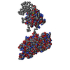
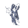
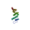
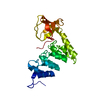
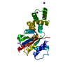
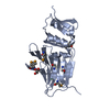
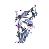
| Title | Crystal structure of the pleckstrin-homology domain of Bcr-Abl in complex with monobody Mb(Bcr-PH_4). | ||||||
 Components Components |
| ||||||
 Keywords Keywords |  SIGNALING PROTEIN / pleckstrin-homology / monobody / Bcr-Abl / phosphoinositide-binding SIGNALING PROTEIN / pleckstrin-homology / monobody / Bcr-Abl / phosphoinositide-binding | ||||||
| Function / homology |  Function and homology information Function and homology informationnegative regulation of respiratory burst / negative regulation of cellular extravasation / negative regulation of macrophage migration / : / negative regulation of blood vessel remodeling / negative regulation of neutrophil degranulation / macrophage migration / neutrophil degranulation / intracellular protein transmembrane transport / renal system process ...negative regulation of respiratory burst / negative regulation of cellular extravasation / negative regulation of macrophage migration / : / negative regulation of blood vessel remodeling / negative regulation of neutrophil degranulation / macrophage migration / neutrophil degranulation / intracellular protein transmembrane transport / renal system process / regulation of vascular permeability / regulation of Rho protein signal transduction / focal adhesion assembly / definitive hemopoiesis / Signaling by cytosolic FGFR1 fusion mutants / activation of GTPase activity / regulation of small GTPase mediated signal transduction / inner ear morphogenesis / small GTPase-mediated signal transduction / RHOB GTPase cycle / RHOC GTPase cycle / CDC42 GTPase cycle / neuromuscular process controlling balance / homeostasis of number of cells / RHOA GTPase cycle / negative regulation of reactive oxygen species metabolic process / RAC2 GTPase cycle / RAC3 GTPase cycle / phagocytosis / positive regulation of phagocytosis / keratinocyte differentiation / RAC1 GTPase cycle / Signaling by FGFR1 in disease / GTPase activator activity / guanyl-nucleotide exchange factor activity / Schaffer collateral - CA1 synapse / brain development / modulation of chemical synaptic transmission / negative regulation of inflammatory response / actin cytoskeleton organization / protein tyrosine kinase activity / cellular response to lipopolysaccharide / dendritic spine / postsynaptic density / non-specific serine/threonine protein kinase / regulation of cell cycle / axon / protein phosphorylation / protein serine kinase activity / protein serine/threonine kinase activity / glutamatergic synapse / signal transduction / protein-containing complex / extracellular exosome / ATP binding / membrane / plasma membrane / cytosolSimilarity search - Function | ||||||
| Biological species |  Homo sapiens (human) Homo sapiens (human)synthetic construct (others) | ||||||
| Method | X-RAY DIFFRACTION / SYNCHROTRON / MOLECULAR REPLACEMENT / Resolution: 1.652 Å | ||||||
 Authors Authors | Reckel, S. / Reynaud, A. / Pojer, F. / Hantschel, O. | ||||||
| Funding support |  Switzerland, 1items Switzerland, 1items
| ||||||
 Citation Citation | Journal: Nat Commun / Year: 2017 Title: Structural and functional dissection of the DH and PH domains of oncogenic Bcr-Abl tyrosine kinase. Authors: Sina Reckel / Charlotte Gehin / Delphine Tardivon / Sandrine Georgeon / Tim Kükenshöner / Frank Löhr / Akiko Koide / Lena Buchner / Alejandro Panjkovich / Aline Reynaud / Sara Pinho / ...Authors: Sina Reckel / Charlotte Gehin / Delphine Tardivon / Sandrine Georgeon / Tim Kükenshöner / Frank Löhr / Akiko Koide / Lena Buchner / Alejandro Panjkovich / Aline Reynaud / Sara Pinho / Barbara Gerig / Dmitri Svergun / Florence Pojer / Peter Güntert / Volker Dötsch / Shohei Koide / Anne-Claude Gavin / Oliver Hantschel /    Abstract: The two isoforms of the Bcr-Abl tyrosine kinase, p210 and p190, are associated with different leukemias and have a dramatically different signaling network, despite similar kinase activity. To ...The two isoforms of the Bcr-Abl tyrosine kinase, p210 and p190, are associated with different leukemias and have a dramatically different signaling network, despite similar kinase activity. To provide a molecular rationale for these observations, we study the Dbl-homology (DH) and Pleckstrin-homology (PH) domains of Bcr-Abl p210, which constitute the only structural differences to p190. Here we report high-resolution structures of the DH and PH domains and characterize conformations of the DH-PH unit in solution. Our structural and functional analyses show no evidence that the DH domain acts as a guanine nucleotide exchange factor, whereas the PH domain binds to various phosphatidylinositol-phosphates. PH-domain mutants alter subcellular localization and result in decreased interactions with p210-selective interaction partners. Hence, the PH domain, but not the DH domain, plays an important role in the formation of the differential p210 and p190 Bcr-Abl signaling networks. | ||||||
| History |
|
- Structure visualization
| Structure viewer | Molecule: MolmilJmol/JSmol |
|---|
- Downloads & links
-Download
| PDBx/mmCIF format | 5oc7.cif.gz | 175.7 KB | Display | PDBx/mmCIF format |
|---|---|---|---|---|
| PDB format | pdb5oc7.ent.gz | 142.2 KB | Display | PDB format |
| PDBx/mmJSON format | 5oc7.json.gz | Tree view | PDBx/mmJSON format | |
| Others |  Other downloads Other downloads |
-Validation report
| Arichive directory | https://data.pdbj.org/pub/pdb/validation_reports/oc/5oc7ftp://data.pdbj.org/pub/pdb/validation_reports/oc/5oc7 | HTTPS FTP |
|---|
-Related structure data
| Related structure data |  5n6rC 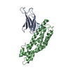 5n7eC 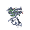 2dfkS S: Starting model for refinement C: citing same article ( |
|---|---|
| Similar structure data |


-Links
 PDBj
PDBj


- Assembly
| Deposited unit | 
| ||||||||
|---|---|---|---|---|---|---|---|---|---|
| 1 | 
| ||||||||
| 2 | 
| ||||||||
| 3 |
| ||||||||
| 4 | 
| ||||||||
| Unit cell |
|
-Components
| #1: Protein | Mass: 15643.062 Da / Num. of mol.: 2 / Mutation: delta 770-829,delta 770-829 Source method: isolated from a genetically manipulated source Source: (gene. exp.) Homo sapiens (human) / Gene: BCR, BCR1, D22S11 / Production host:  Escherichia coli BL21(DE3) (bacteria) Escherichia coli BL21(DE3) (bacteria)References: UniProt: P11274, non-specific serine/threonine protein kinase#2: Protein | Mass: 9632.773 Da / Num. of mol.: 2 Source method: isolated from a genetically manipulated source Source: (gene. exp.) synthetic construct (others) / Production host: Escherichia coli BL21(DE3) (bacteria)#3: Chemical | ChemComp-GOL / Glycerol  Mass: 92.094 Da / Num. of mol.: 7 / Source method: obtained synthetically / Formula: C3H8O3 Mass: 92.094 Da / Num. of mol.: 7 / Source method: obtained synthetically / Formula: C3H8O3#4: Chemical | ChemComp-IP2 / |   Mass: 340.116 Da / Num. of mol.: 1 / Source method: obtained synthetically / Formula: C6H14O12P2 / Feature type: SUBJECT OF INVESTIGATION Mass: 340.116 Da / Num. of mol.: 1 / Source method: obtained synthetically / Formula: C6H14O12P2 / Feature type: SUBJECT OF INVESTIGATION#5: Water | ChemComp-HOH / | Water Mass: 18.015 Da / Num. of mol.: 253 / Source method: isolated from a natural source / Formula: H2O Mass: 18.015 Da / Num. of mol.: 253 / Source method: isolated from a natural source / Formula: H2O |
|---|
-Experimental details
-Experiment
| Experiment | Method: X-RAY DIFFRACTION / Number of used crystals: 1 |
|---|
- Sample preparation
| Crystal | Density Matthews: 2.47 Å3/Da / Density % sol: 50 % |
|---|---|
| Crystal grow | Temperature: 291 K / Method: vapor diffusion, sitting drop Details: 0.1 M Potassium thiocyanate, 30% (w/v) PEG MME 2000 |
-Data collection
| Diffraction | Mean temperature: 100 K |
|---|---|
| Diffraction source | Source: SYNCHROTRON / Site: SLS / Beamline: X06DA / Wavelength: 0.99987 Å |
| Detector | Type: DECTRIS PILATUS 2M / Detector: PIXEL / Date: May 19, 2017 |
| Radiation | Protocol: SINGLE WAVELENGTH / Monochromatic (M) / Laue (L): M / Scattering type: x-ray |
| Radiation wavelength | Wavelength: 0.99987 Å / Relative weight: 1 |
| Reflection | Resolution: 1.652→33.68 Å / Num. obs: 49951 / % possible obs: 97.13 % / Redundancy: 4.899 % / Rmerge(I) obs: 0.03096 / Rpim(I) all: 0.01554 / Rrim(I) all: 0.03473 / Net I/σ(I): 23.01 |
| Reflection shell | Resolution: 1.652→1.711 Å / Rmerge(I) obs: 0.6334 / Num. unique obs: 4899 / Rpim(I) all: 0.3281 / Rrim(I) all: 0.7159 / % possible all: 96.1 |
- Processing
| Software |
| |||||||||||||||||||||||||||||||||||||||||||||||||||||||||||||||||||||||||||||||||||||||||||||||||||||||||||||||||||||||||||||||||||||||||||||||||||||||||||||||||||||||||||||||||||||||||||||||||||||||||||
|---|---|---|---|---|---|---|---|---|---|---|---|---|---|---|---|---|---|---|---|---|---|---|---|---|---|---|---|---|---|---|---|---|---|---|---|---|---|---|---|---|---|---|---|---|---|---|---|---|---|---|---|---|---|---|---|---|---|---|---|---|---|---|---|---|---|---|---|---|---|---|---|---|---|---|---|---|---|---|---|---|---|---|---|---|---|---|---|---|---|---|---|---|---|---|---|---|---|---|---|---|---|---|---|---|---|---|---|---|---|---|---|---|---|---|---|---|---|---|---|---|---|---|---|---|---|---|---|---|---|---|---|---|---|---|---|---|---|---|---|---|---|---|---|---|---|---|---|---|---|---|---|---|---|---|---|---|---|---|---|---|---|---|---|---|---|---|---|---|---|---|---|---|---|---|---|---|---|---|---|---|---|---|---|---|---|---|---|---|---|---|---|---|---|---|---|---|---|---|---|---|---|---|---|---|
| Refinement | Method to determine structure: MOLECULAR REPLACEMENT Starting model: 2DFK Resolution: 1.652→33.68 Å / SU ML: 0.2 / Cross valid method: NONE / σ(F): 1.97 / Phase error: 21.66
| |||||||||||||||||||||||||||||||||||||||||||||||||||||||||||||||||||||||||||||||||||||||||||||||||||||||||||||||||||||||||||||||||||||||||||||||||||||||||||||||||||||||||||||||||||||||||||||||||||||||||||
| Solvent computation | Shrinkage radii: 0.9 Å / VDW probe radii: 1.11 Å | |||||||||||||||||||||||||||||||||||||||||||||||||||||||||||||||||||||||||||||||||||||||||||||||||||||||||||||||||||||||||||||||||||||||||||||||||||||||||||||||||||||||||||||||||||||||||||||||||||||||||||
| Refinement step | Cycle: LAST / Resolution: 1.652→33.68 Å
| |||||||||||||||||||||||||||||||||||||||||||||||||||||||||||||||||||||||||||||||||||||||||||||||||||||||||||||||||||||||||||||||||||||||||||||||||||||||||||||||||||||||||||||||||||||||||||||||||||||||||||
| Refine LS restraints |
| |||||||||||||||||||||||||||||||||||||||||||||||||||||||||||||||||||||||||||||||||||||||||||||||||||||||||||||||||||||||||||||||||||||||||||||||||||||||||||||||||||||||||||||||||||||||||||||||||||||||||||
| LS refinement shell |
|
