 Movie
Movie Controller

[English] 日本語
 Yorodumi
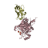
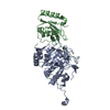
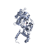
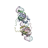
- PDB-4z2b: The structure of human PDE12 residues 161-609 in complex with GSK... -
+ Open data

- Basic information
| Entry | Database: PDB / ID: 4z2b | ||||||
|---|---|---|---|---|---|---|---|
| Title | The structure of human PDE12 residues 161-609 in complex with GSK3036342A | ||||||
 Components Components | 2',5'-phosphodiesterase 12 | ||||||
 Keywords Keywords | Hydrolase/Hydrolase Inhibitor / PDE12 2'-5'A EEP nuclease Inhibitor complex / Hydrolase-Hydrolase Inhibitor complex | ||||||
| Function / homology |  Function and homology information Function and homology informationoligoribonucleotidase activity / mitochondrial mRNA catabolic process / regulation of mitochondrial mRNA stability /  poly(A)-specific ribonuclease / poly(A)-specific ribonuclease activity / cellular response to interferon-alpha / nucleic acid metabolic process / OAS antiviral response / Hydrolases; Acting on ester bonds; Phosphoric-diester hydrolases / cellular response to dsRNA ...oligoribonucleotidase activity / mitochondrial mRNA catabolic process / regulation of mitochondrial mRNA stability / poly(A)-specific ribonuclease / poly(A)-specific ribonuclease activity / cellular response to interferon-alpha / nucleic acid metabolic process / OAS antiviral response / Hydrolases; Acting on ester bonds; Phosphoric-diester hydrolases / cellular response to dsRNA / nuclear-transcribed mRNA catabolic process, deadenylation-dependent decay / exonuclease activity / antiviral innate immune response / positive regulation of viral genome replication / mRNA processing / cellular response to type II interferon / 3'-5'-RNA exonuclease activity / defense response to virus / mitochondrial matrix / mitochondrion / metal ion binding / cytosol poly(A)-specific ribonuclease / poly(A)-specific ribonuclease activity / cellular response to interferon-alpha / nucleic acid metabolic process / OAS antiviral response / Hydrolases; Acting on ester bonds; Phosphoric-diester hydrolases / cellular response to dsRNA ...oligoribonucleotidase activity / mitochondrial mRNA catabolic process / regulation of mitochondrial mRNA stability / poly(A)-specific ribonuclease / poly(A)-specific ribonuclease activity / cellular response to interferon-alpha / nucleic acid metabolic process / OAS antiviral response / Hydrolases; Acting on ester bonds; Phosphoric-diester hydrolases / cellular response to dsRNA / nuclear-transcribed mRNA catabolic process, deadenylation-dependent decay / exonuclease activity / antiviral innate immune response / positive regulation of viral genome replication / mRNA processing / cellular response to type II interferon / 3'-5'-RNA exonuclease activity / defense response to virus / mitochondrial matrix / mitochondrion / metal ion binding / cytosolSimilarity search - Function | ||||||
| Biological species |  Homo sapiens (human) Homo sapiens (human) | ||||||
| Method | X-RAY DIFFRACTION / MOLECULAR REPLACEMENT / molecular replacement / Resolution: 1.8 Å | ||||||
 Authors Authors | Nolte, R.T. / Wisely, B. / Wang, L. / Wood, E.R. | ||||||
 Citation Citation | Journal: J.Biol.Chem. / Year: 2015 Title: The Role of Phosphodiesterase 12 (PDE12) as a Negative Regulator of the Innate Immune Response and the Discovery of Antiviral Inhibitors. Authors: Wood, E.R. / Bledsoe, R. / Chai, J. / Daka, P. / Deng, H. / Ding, Y. / Harris-Gurley, S. / Kryn, L.H. / Nartey, E. / Nichols, J. / Nolte, R.T. / Prabhu, N. / Rise, C. / Sheahan, T. / ...Authors: Wood, E.R. / Bledsoe, R. / Chai, J. / Daka, P. / Deng, H. / Ding, Y. / Harris-Gurley, S. / Kryn, L.H. / Nartey, E. / Nichols, J. / Nolte, R.T. / Prabhu, N. / Rise, C. / Sheahan, T. / Shotwell, J.B. / Smith, D. / Tai, V. / Taylor, J.D. / Tomberlin, G. / Wang, L. / Wisely, B. / You, S. / Xia, B. / Dickson, H. | ||||||
| History |
|
- Structure visualization
| Structure viewer | Molecule: MolmilJmol/JSmol |
|---|
- Downloads & links
-Download
| PDBx/mmCIF format | 4z2b.cif.gz | 120.4 KB | Display | PDBx/mmCIF format |
|---|---|---|---|---|
| PDB format | pdb4z2b.ent.gz | 87.6 KB | Display | PDB format |
| PDBx/mmJSON format | 4z2b.json.gz | Tree view | PDBx/mmJSON format | |
| Others |  Other downloads Other downloads |
-Validation report
| Arichive directory | https://data.pdbj.org/pub/pdb/validation_reports/z2/4z2bftp://data.pdbj.org/pub/pdb/validation_reports/z2/4z2b | HTTPS FTP |
|---|
-Related structure data
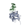
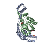
| Related structure data | 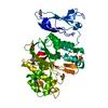 4z0vC 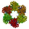 3ng0S C: citing same article ( S: Starting model for refinement |
|---|---|
| Similar structure data |


-Links
 PDBj
PDBj


- Assembly
| Deposited unit | 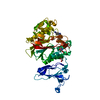
| ||||||||
|---|---|---|---|---|---|---|---|---|---|
| 1 |
| ||||||||
| Unit cell |
|
-Components
-Protein , 1 types, 1 molecules A
| #1: Protein | Mass: 51137.672 Da / Num. of mol.: 1 / Fragment: UNP residues 155-609 Source method: isolated from a genetically manipulated source Details: Tev cloning tag on N-terminus / Source: (gene. exp.) Homo sapiens (human) / Gene: PDE12 / Production host:  Escherichia coli (E. coli) / Strain (production host): BL21 / Variant (production host): DE3 Escherichia coli (E. coli) / Strain (production host): BL21 / Variant (production host): DE3References: UniProt: Q6L8Q7, Hydrolases; Acting on ester bonds; Phosphoric-diester hydrolases, poly(A)-specific ribonuclease |
|---|
-Non-polymers , 6 types, 468 molecules 


| #2: Chemical | ChemComp-SO4 / Sulfate Mass: 96.063 Da / Num. of mol.: 1 / Source method: obtained synthetically / Formula: SO4 Mass: 96.063 Da / Num. of mol.: 1 / Source method: obtained synthetically / Formula: SO4 | ||||||||
|---|---|---|---|---|---|---|---|---|---|
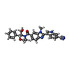
| #3: Chemical | Ethylene glycol Mass: 62.068 Da / Num. of mol.: 2 / Source method: obtained synthetically / Formula: C2H6O2 Mass: 62.068 Da / Num. of mol.: 2 / Source method: obtained synthetically / Formula: C2H6O2#4: Chemical | ChemComp-TRS / | Tris Mass: 122.143 Da / Num. of mol.: 1 / Source method: obtained synthetically / Formula: C4H12NO3 / Comment: pH buffer*YM Mass: 122.143 Da / Num. of mol.: 1 / Source method: obtained synthetically / Formula: C4H12NO3 / Comment: pH buffer*YM#5: Chemical | ChemComp-4LC / |  Mass: 491.541 Da / Num. of mol.: 1 / Source method: obtained synthetically / Formula: C29H25N5O3 Mass: 491.541 Da / Num. of mol.: 1 / Source method: obtained synthetically / Formula: C29H25N5O3#6: Chemical | ChemComp-MG / |  Mass: 24.305 Da / Num. of mol.: 1 / Source method: obtained synthetically / Formula: Mg Mass: 24.305 Da / Num. of mol.: 1 / Source method: obtained synthetically / Formula: Mg#7: Water | ChemComp-HOH / | WaterMass: 18.015 Da / Num. of mol.: 462 / Source method: isolated from a natural source / Formula: H2O |
-Experimental details
-Experiment
| Experiment | Method: X-RAY DIFFRACTION / Number of used crystals: 1 |
|---|
- Sample preparation
| Crystal | Density Matthews: 2.32 Å3/Da / Density % sol: 47.07 % |
|---|---|
| Crystal grow | Temperature: 295 K / Method: vapor diffusion, sitting drop / pH: 8.5 / Details: 0.1M Tris Ph 8.5 15% Peg20000 25% Ethylene Glycol |
-Data collection
| Diffraction | Mean temperature: 100 K | |||||||||||||||||||||||||||||||||||||||||||||||||||||||||||||||||||||||||||||||||||||||||||||||||||||||||||||||||||||||||||||||||||||||||||||||||||||||||||||||||||||||||||||||||||||||||||||
|---|---|---|---|---|---|---|---|---|---|---|---|---|---|---|---|---|---|---|---|---|---|---|---|---|---|---|---|---|---|---|---|---|---|---|---|---|---|---|---|---|---|---|---|---|---|---|---|---|---|---|---|---|---|---|---|---|---|---|---|---|---|---|---|---|---|---|---|---|---|---|---|---|---|---|---|---|---|---|---|---|---|---|---|---|---|---|---|---|---|---|---|---|---|---|---|---|---|---|---|---|---|---|---|---|---|---|---|---|---|---|---|---|---|---|---|---|---|---|---|---|---|---|---|---|---|---|---|---|---|---|---|---|---|---|---|---|---|---|---|---|---|---|---|---|---|---|---|---|---|---|---|---|---|---|---|---|---|---|---|---|---|---|---|---|---|---|---|---|---|---|---|---|---|---|---|---|---|---|---|---|---|---|---|---|---|---|---|---|---|---|
| Diffraction source | Source: ROTATING ANODE / Type: RIGAKU FR-E+ SUPERBRIGHT / Wavelength: 1.54 Å | |||||||||||||||||||||||||||||||||||||||||||||||||||||||||||||||||||||||||||||||||||||||||||||||||||||||||||||||||||||||||||||||||||||||||||||||||||||||||||||||||||||||||||||||||||||||||||||
| Detector | Type: RIGAKU SATURN 944+ / Detector: CCD / Date: Sep 12, 2012 | |||||||||||||||||||||||||||||||||||||||||||||||||||||||||||||||||||||||||||||||||||||||||||||||||||||||||||||||||||||||||||||||||||||||||||||||||||||||||||||||||||||||||||||||||||||||||||||
| Radiation | Monochromator: Multilayer optics / Protocol: SINGLE WAVELENGTH / Monochromatic (M) / Laue (L): M / Scattering type: x-ray | |||||||||||||||||||||||||||||||||||||||||||||||||||||||||||||||||||||||||||||||||||||||||||||||||||||||||||||||||||||||||||||||||||||||||||||||||||||||||||||||||||||||||||||||||||||||||||||
| Radiation wavelength | Wavelength: 1.54 Å / Relative weight: 1 | |||||||||||||||||||||||||||||||||||||||||||||||||||||||||||||||||||||||||||||||||||||||||||||||||||||||||||||||||||||||||||||||||||||||||||||||||||||||||||||||||||||||||||||||||||||||||||||
| Reflection | Redundancy: 4.8 % / Number: 194359 / Rmerge(I) obs: 0.054 / Χ2: 0.94 / D res high: 1.8 Å / D res low: 50 Å / Num. obs: 40745 / % possible obs: 91.9 | |||||||||||||||||||||||||||||||||||||||||||||||||||||||||||||||||||||||||||||||||||||||||||||||||||||||||||||||||||||||||||||||||||||||||||||||||||||||||||||||||||||||||||||||||||||||||||||
| Diffraction reflection shell |
| |||||||||||||||||||||||||||||||||||||||||||||||||||||||||||||||||||||||||||||||||||||||||||||||||||||||||||||||||||||||||||||||||||||||||||||||||||||||||||||||||||||||||||||||||||||||||||||
| Reflection | Resolution: 1.8→50 Å / Num. obs: 40745 / % possible obs: 91.9 % / Redundancy: 4.8 % / Biso Wilson estimate: 20.62 Å2 / Rmerge(I) obs: 0.054 / Net I/σ(I): 15.2 | |||||||||||||||||||||||||||||||||||||||||||||||||||||||||||||||||||||||||||||||||||||||||||||||||||||||||||||||||||||||||||||||||||||||||||||||||||||||||||||||||||||||||||||||||||||||||||||
| Reflection shell | Diffraction-ID: 1 / Rejects: 0
|
-Phasing
| Phasing | Method: molecular replacement | |||||||||
|---|---|---|---|---|---|---|---|---|---|---|
| Phasing MR |
|
- Processing
| Software |
| ||||||||||||||||||||||||||||||||||||||||||||||||||||||||||||||||||||||||||||||||||||||||||||||||||||||||||||||||||
|---|---|---|---|---|---|---|---|---|---|---|---|---|---|---|---|---|---|---|---|---|---|---|---|---|---|---|---|---|---|---|---|---|---|---|---|---|---|---|---|---|---|---|---|---|---|---|---|---|---|---|---|---|---|---|---|---|---|---|---|---|---|---|---|---|---|---|---|---|---|---|---|---|---|---|---|---|---|---|---|---|---|---|---|---|---|---|---|---|---|---|---|---|---|---|---|---|---|---|---|---|---|---|---|---|---|---|---|---|---|---|---|---|---|---|---|
| Refinement | Method to determine structure: MOLECULAR REPLACEMENT Starting model: 3NG0 Resolution: 1.8→27.3 Å / Cor.coef. Fo:Fc: 0.9524 / Cor.coef. Fo:Fc free: 0.9254 / SU R Cruickshank DPI: 0.141 / Cross valid method: THROUGHOUT / σ(F): 0 / SU R Blow DPI: 0.153 / SU Rfree Blow DPI: 0.135 / SU Rfree Cruickshank DPI: 0.13
| ||||||||||||||||||||||||||||||||||||||||||||||||||||||||||||||||||||||||||||||||||||||||||||||||||||||||||||||||||
| Displacement parameters | Biso mean: 21.79 Å2
| ||||||||||||||||||||||||||||||||||||||||||||||||||||||||||||||||||||||||||||||||||||||||||||||||||||||||||||||||||
| Refine analyze | Luzzati coordinate error obs: 0.201 Å | ||||||||||||||||||||||||||||||||||||||||||||||||||||||||||||||||||||||||||||||||||||||||||||||||||||||||||||||||||
| Refinement step | Cycle: LAST / Resolution: 1.8→27.3 Å
| ||||||||||||||||||||||||||||||||||||||||||||||||||||||||||||||||||||||||||||||||||||||||||||||||||||||||||||||||||
| Refine LS restraints |
| ||||||||||||||||||||||||||||||||||||||||||||||||||||||||||||||||||||||||||||||||||||||||||||||||||||||||||||||||||
| LS refinement shell | Resolution: 1.8→1.85 Å / Total num. of bins used: 20
|
