 Movie
Movie Controller


+ Open data

- Basic information
| Entry | Database: PDB / ID: 6bzg | ||||||
|---|---|---|---|---|---|---|---|
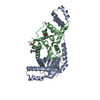
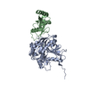
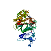
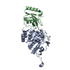
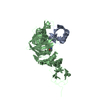
| Title | Structure of S. cerevisiae Zip2:Spo16 complex, P212121 form | ||||||
 Components Components |
| ||||||
 Keywords Keywords |  DNA BINDING PROTEIN / XPF-ERCC1 / Meiosis / Recombination DNA BINDING PROTEIN / XPF-ERCC1 / Meiosis / Recombination | ||||||
| Function / homology |  Function and homology informationregulation of synaptonemal complex assembly / synapsis initiation complex / regulation of reciprocal meiotic recombination / DNA secondary structure binding / ascospore formation / synaptonemal complex assembly / homologous chromosome pairing at meiosis / synaptonemal complex / positive regulation of protein sumoylation / homologous recombination ...regulation of synaptonemal complex assembly / synapsis initiation complex / regulation of reciprocal meiotic recombination / DNA secondary structure binding / ascospore formation / synaptonemal complex assembly / homologous chromosome pairing at meiosis / synaptonemal complex / positive regulation of protein sumoylation / homologous recombination / reciprocal meiotic recombination / protein sumoylation / condensed nuclear chromosome / nucleotide binding / mitochondrion Function and homology informationregulation of synaptonemal complex assembly / synapsis initiation complex / regulation of reciprocal meiotic recombination / DNA secondary structure binding / ascospore formation / synaptonemal complex assembly / homologous chromosome pairing at meiosis / synaptonemal complex / positive regulation of protein sumoylation / homologous recombination ...regulation of synaptonemal complex assembly / synapsis initiation complex / regulation of reciprocal meiotic recombination / DNA secondary structure binding / ascospore formation / synaptonemal complex assembly / homologous chromosome pairing at meiosis / synaptonemal complex / positive regulation of protein sumoylation / homologous recombination / reciprocal meiotic recombination / protein sumoylation / condensed nuclear chromosome / nucleotide binding / mitochondrionSimilarity search - Function | ||||||
| Biological species |  Saccharomyces cerevisiae (brewer's yeast) Saccharomyces cerevisiae (brewer's yeast) | ||||||
| Method | X-RAY DIFFRACTION / SYNCHROTRON / SAD / Resolution: 2.13 Å | ||||||
 Authors Authors | Arora, K. / Corbett, K.D. | ||||||
| Funding support |  United States, 1items United States, 1items
| ||||||
 Citation Citation | Journal: Nucleic Acids Res. / Year: 2019 Title: The conserved XPF:ERCC1-like Zip2:Spo16 complex controls meiotic crossover formation through structure-specific DNA binding. Authors: Arora, K. / Corbett, K.D. #1: Journal: Biorxiv / Year: 2018Title: Structure of Zip2:Spo16, a conserved XPF:ERCC1-like complex critical for meiotic crossover formation Authors: Arora, K. / Corbett, K.D. | ||||||
| History |
|
- Structure visualization
| Structure viewer | Molecule: MolmilJmol/JSmol |
|---|
- Downloads & links
-Download
| PDBx/mmCIF format | 6bzg.cif.gz | 178.9 KB | Display | PDBx/mmCIF format |
|---|---|---|---|---|
| PDB format | pdb6bzg.ent.gz | 142.4 KB | Display | PDB format |
| PDBx/mmJSON format | 6bzg.json.gz | Tree view | PDBx/mmJSON format | |
| Others |  Other downloads Other downloads |
-Validation report
| Arichive directory | https://data.pdbj.org/pub/pdb/validation_reports/bz/6bzgftp://data.pdbj.org/pub/pdb/validation_reports/bz/6bzg | HTTPS FTP |
|---|
-Related structure data
| Related structure data |  6bzfC C: citing same article ( |
|---|---|
| Similar structure data | |
| Experimental dataset #1 | Data reference: 10.15785/SBGRID/539 / Data set type: diffraction image data |
| Experimental dataset #2 | Data reference: 10.15785/SBGRID/540 / Data set type: diffraction image data |


-Links
 PDBj
PDBj- Assembly
| Deposited unit | 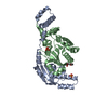
| ||||||||
|---|---|---|---|---|---|---|---|---|---|
| 1 |
| ||||||||
| Unit cell |
|
-Components
| #1: Protein | Mass: 24345.135 Da / Num. of mol.: 1 / Mutation: K641A, E642A, K643A Source method: isolated from a genetically manipulated source Source: (gene. exp.) Saccharomyces cerevisiae (brewer's yeast)Gene: ZIP2 / Production host:  Escherichia coli (E. coli) / References: UniProt: P53061 Escherichia coli (E. coli) / References: UniProt: P53061 | ||||
|---|---|---|---|---|---|
| #2: Protein | Mass: 23023.074 Da / Num. of mol.: 1 Source method: isolated from a genetically manipulated source Source: (gene. exp.) Saccharomyces cerevisiae (brewer's yeast)Gene: SPO16 / Production host: Escherichia coli (E. coli) / References: UniProt: P17122 | ||||
| #3: Chemical | Sulfate  Mass: 96.063 Da / Num. of mol.: 2 / Source method: obtained synthetically / Formula: SO4 Mass: 96.063 Da / Num. of mol.: 2 / Source method: obtained synthetically / Formula: SO4#4: Chemical | ChemComp-P6G / | Polyethylene glycol  Mass: 282.331 Da / Num. of mol.: 1 / Source method: obtained synthetically / Formula: C12H26O7 / Comment: precipitant*YM Mass: 282.331 Da / Num. of mol.: 1 / Source method: obtained synthetically / Formula: C12H26O7 / Comment: precipitant*YM#5: Water | ChemComp-HOH / | Water Mass: 18.015 Da / Num. of mol.: 74 / Source method: isolated from a natural source / Formula: H2O Mass: 18.015 Da / Num. of mol.: 74 / Source method: isolated from a natural source / Formula: H2O |
-Experimental details
-Experiment
| Experiment | Method: X-RAY DIFFRACTION / Number of used crystals: 1 |
|---|
- Sample preparation
| Crystal | Density Matthews: 2.59 Å3/Da / Density % sol: 52.48 % |
|---|---|
| Crystal grow | Temperature: 293 K / Method: vapor diffusion, hanging drop / pH: 5.5 Details: 100 mM Bis Tris pH 5.5, 200 mM Ammonium sulfate, 15% PEG-3350 |
-Data collection
| Diffraction | Mean temperature: 100 K |
|---|---|
| Diffraction source | Source: SYNCHROTRON / Site: APS / Beamline: 24-ID-C / Wavelength: 0.9792 Å |
| Detector | Type: DECTRIS PILATUS3 6M / Detector: PIXEL / Date: Jun 15, 2017 |
| Radiation | Protocol: SINGLE WAVELENGTH / Monochromatic (M) / Laue (L): M / Scattering type: x-ray |
| Radiation wavelength | Wavelength: 0.9792 Å / Relative weight: 1 |
| Reflection | Resolution: 2.13→100 Å / Num. obs: 28219 / % possible obs: 99.8 % / Redundancy: 6.3 % / CC1/2: 0.987 / Rsym value: 0.207 / Net I/σ(I): 5.2 |
| Reflection shell | Resolution: 2.13→2.19 Å / Num. unique obs: 15501 / CC1/2: 0.484 / Rsym value: 2.074 |
- Processing
| Software |
| ||||||||||||||||||||||||||||||||||||||||||||||||||||||||||||||||||||||||||||||||||||||||||||||||||||||||||||||||||||||||||||||||||||||||||||
|---|---|---|---|---|---|---|---|---|---|---|---|---|---|---|---|---|---|---|---|---|---|---|---|---|---|---|---|---|---|---|---|---|---|---|---|---|---|---|---|---|---|---|---|---|---|---|---|---|---|---|---|---|---|---|---|---|---|---|---|---|---|---|---|---|---|---|---|---|---|---|---|---|---|---|---|---|---|---|---|---|---|---|---|---|---|---|---|---|---|---|---|---|---|---|---|---|---|---|---|---|---|---|---|---|---|---|---|---|---|---|---|---|---|---|---|---|---|---|---|---|---|---|---|---|---|---|---|---|---|---|---|---|---|---|---|---|---|---|---|---|---|
| Refinement | Method to determine structure: SAD / Resolution: 2.13→69.809 Å / SU ML: 0.4 / Cross valid method: FREE R-VALUE / σ(F): 1.2 / Phase error: 32.61
| ||||||||||||||||||||||||||||||||||||||||||||||||||||||||||||||||||||||||||||||||||||||||||||||||||||||||||||||||||||||||||||||||||||||||||||
| Solvent computation | Shrinkage radii: 0.9 Å / VDW probe radii: 1.11 Å | ||||||||||||||||||||||||||||||||||||||||||||||||||||||||||||||||||||||||||||||||||||||||||||||||||||||||||||||||||||||||||||||||||||||||||||
| Refinement step | Cycle: LAST / Resolution: 2.13→69.809 Å
| ||||||||||||||||||||||||||||||||||||||||||||||||||||||||||||||||||||||||||||||||||||||||||||||||||||||||||||||||||||||||||||||||||||||||||||
| Refine LS restraints |
| ||||||||||||||||||||||||||||||||||||||||||||||||||||||||||||||||||||||||||||||||||||||||||||||||||||||||||||||||||||||||||||||||||||||||||||
| LS refinement shell |
| ||||||||||||||||||||||||||||||||||||||||||||||||||||||||||||||||||||||||||||||||||||||||||||||||||||||||||||||||||||||||||||||||||||||||||||
| Refinement TLS params. | Method: refined / Refine-ID: X-RAY DIFFRACTION
| ||||||||||||||||||||||||||||||||||||||||||||||||||||||||||||||||||||||||||||||||||||||||||||||||||||||||||||||||||||||||||||||||||||||||||||
| Refinement TLS group |
|
